![]()
ANTIOXIDANTE
EFFECT IN VITRO OF THE HOMEOPATHIC MEDICINE ARSENICUM ALBUM, CUPRUM
METALLICUM MANGANUM AND ZINCUM METALLICUM.
CELSO FERNANDES
BATELLO
batello@batello.med.brDissertation presented as part of the demanded
requirements to obtain the title of MA in Homeopathy by FACIS – IBEHE
Tutor: Professor Dr. Vânia d´ Almeida
SÃO PAULO – 2002
FACIS – HEALTH SCIENCE COLLEGE OF SÃO PAULO
IBEHE – CENTER OF POST-GRADUATION IN HOMEOPATHY
Português.
DEDICATION
I dedicate this work, first and above all, to God,
with honor and glory.
To my parents, Clarisse and Oscar Batello for the love, affection and
dedication.
And to my wonderful children Caio Márcio and Marcella Helena, wheat and
honey of my existence.
THANKS
I thank Professor Dr. Vânia d´Almeida by the
scientific spirit and orientation for the development of this work.To Professor Dr. Ana Maria Martins by accepting the
challenge!To the Health Science College in São Paulo and
Brazilian Institute of Homeopathic Studies an others, by being pioneer and
brave enough to create, maybe, the first MA Homeopathy in the world.
Brazil needs a lot this kind of initiative.To São Paulo Federal University.
To the professors, employees and friends from the
Health Science College of São Paulo and from the Brazilian Institute of
Homeopathic Studies and others, by affection and dedication that always
gave to my person.To the graduating friends from the first MA in
Homeopathy of the Health Science College in São Paulo that became my
friends, and also by the knowledge they transmitted.To Dr. Marcelo Pustiglione, Homeopathy Professor by the
friendship and deep knowledge in Medicine mainly in Homeopathy, that
transmitted and coordinated this Ma eight much competence.To the Professor Dr(s) from the Health Science College
in São Paulo, Maria de Lourdes Brunini,Célia Maria Piva Sena, José
Carlos Tavares, Leone Villario Bonamin, Elisabeth Teresa Brunini
Sbardelini, Jorge Camilo Flório, by the effort, affection and dedication.To the psychobiology Department of the Escola Paulista
de Medicina, especially to Professor Sérgio Tufik, for the collaboration
in obtunding the animals.To the pharmacist graduating in Homeopathy in the
Health Science College in São Paulo, Dr. Edson Godoy for the essential
help in the rigorous preparation of the homeopathic Medicine.To the professors, students and employees from the
Medical Genetic Center of the Pediatry and Morphology Department of the
São Paulo Federal University, especially to Carolina do Amaral Terzi,
that collaborated the experimental part of this work.
ABSTRACT
This dissertation, with the support of a theoretical
practical foundation, presents the scenario where is inserted the
problematic in question; The Antioxidant Effect in vitro of
Homeopathic Medicines Arsenicum album, Cuprum metallicum, Manganum and
Zincum metallicum.It is demonstrated in the first chapters, of
theoretical bibliographical substantiation, the Homeopathy and
Oligotherapy as therapeutics techniques, as well as the importance of
oxidation phenomena for a better comprehension of the organic phenomena,
mainly in the genesis of many diseases. It is also experimentally
demonstrated the homeopathic medicines antioxidant action in different
dilutions in comparison with melatonin in various concentrations over the
lipidic peroxidation in homogenate of mice brains measured though the
malondialdehyde dosage obtained through the absorbency technique.For the analysis of the results the Kruskal-Wallys and
Dunn´s Multiple Comparisons tests were realized, that revealed
significant differences among the experimented groups.It was verified a greater lipidic peroxidation
inhibiting effect with melatonin 1M, followed by melatonin 0.5M, Cuprum
metallicum C12, Cuprum Metallicum C30, Arsenicum album C30, melatonine
0.24M, Manganum C30 and Arsenicum album C12.It was proved that the melatonin has an in vitro
lipidic peroxidation inhibiting effect, and so being adopted as reference.
However the new fact arises from the observation of the significant
lipidic peroxidation inhibition obtained with the usage of Homeopathic
medicines, sometimes with dilutions that supersed the Avogadro number, as
in the cases of Cuprum metallicum C30, Arsenicum album C30 and Manganum
C30 in decreasing action order.This work calls the attention for the possibility of
existence of an antioxidant mechanism action of homeopathic medicine
different from the know cause effect relationship.
SUMMARY
CHAPTER
1.INTRODUCTION
1.1. Medicine History
1.1.1 Homeopathy History11
1.1.2 Hahnemann, Homeopathy History Father
1.1.3 Homeopathy History in Brazil
1.1.4 Homeopathy and Allopathy
1.2. Medicine
1.2.1. Homeopathic Medicine
1.3 Origin of the homeopathic medicine
1.4 Medicine experimented in the healthy man
1.5 Homeopathic Medicine Action
1.6 Homeopathic Medicine Experimentation
1.6.1 Zinc
1.6.2 Zincum Metallicum
1.6.3 Copper
1.6.4 Cuprum Metallicum
1.6.5 Arsenic
1.6.6 Arsenicum álbum
1.6.7 Manganese
1.6.8 Manganum
1.7 Reactive Species of Oxygen
1.7.1 Oxygen Origin
1.7.2 Oxygen metabolism
1.7.3 Free Radical
1.7.4 Hydrogen Peroxide
1.7.5 Fenton Reaction
1.7.6 Haber – Weiss Reaction
1.8 Other Physiological Conditions
1.9 Reactive Species of Oxygen and Antioxidant systems
1.10 Reactive Species Oxygen Formation
1.11 RSO Action in the biological systems
1.11.1 Ischemia and reperfusion
1.11.2 Neurologic Diseases
1.11.3 Lipidic Peroxidation
1.12 Antioxidant mechanisms
1.13 Antioxidant Defense
1.14 No-enzymatic antioxidant
1.14.1 The Melatonin
1.14.2 Minerals
1.14.3Enzymatic Antioxidant
1.14.4 Enzym
1.14.4.1 Dismutase Superoxid
1.14.4.1.1 Dismutase Superoxid dependent on copper – zinc
1.14.4.1.2 Dismutase Superoxid dependent on manganese
1.14.4.1.3 Dismutase Superoxid extra–cellular
1.14.4.1.4 Particularities
1.14.4.2 Glutathione Peroxidase
1.14.4.3 Catalase
1.14.4.3.1 Interaction
1.15 Oligoelements
1.15.1 quantitative Difference
1.15.2 Cofactors
1.15.3 Enzymatic Process2. GOALS
3. MATERIALS AND METHODS
3.1 Materials
3.1.1 Animals
3.1.2 Reagents and equipment’s
3.2 Methods
3.2.1 Research Methodology
3.2.2. Statistic Method4. RESULTS
5. DISCUSSION
6. CONCLUSION
7. ABREVIATIONS AND SYMBOLS
8. BIBLIOGRAPHY
INDEX
CHART/PICTURE
CHART 1: Oxygen Reactive Species
CHART 2: Oxygen Reactive Species and antioxidants
CHART 3: Melatonin Concentration used in the experiment
CHART 4: Medicine used and their respective potencies
CHART 5: Results got in the analysis of multiple
comparisons by DunnCHART 6: Significance Level of the Different Experimental groups got by
Dunn´s analysis of multiple comparisonsCHART 7: Percentage of lipidic peroxidation in homogenate of mice’s
brain trying the homeopathic medicine Cuprum Metallicum, Zincum
Metallicum, Manganum and Arsenicum album, Comparatively to themelation M
0,125 molarGRAPHIC 1: Inhibition percentage of the
lipoperoxidation.
1.
INTRODUCTIONThis work was idealized starting by the presupposition
that, if the homeopathic medicine possesses therapeutic and preventive
action, maybe, part of this action comes from an antioxidant action.Based on Hahnneman, paragraph 20, justificative for the
experimentation:“We aren’t abees to find out this immaterial
power that is found latent in the intimus essence of the medicine only
with the reason’s efforts. Just by the experience (experimentation), we
can note clearly the phenomena that it provokes when it acts on the
healthy organism (experimentation in the healthy man)” (Pustiglione,
2001).Analogously, experimentation will be made, but in
vitro, to verify the possible antioxidant of mice’s brain, comparatively
to melatonin, specifically in the lipidic peroxidation. As a measure of
this antioxidant action, the malondialdehyde (MDA) will be dosed,
resulting from the lipidic peroxidion reactions.We intend to show by experience, in vitro, the
homeopathic medicine action in a specific stage of the cascade of reactive
species of oxygen, the lipidic perioxidation, trying to understand,
through the laboratory, and by inference, the effects of these medicines
in the healthy man.We even intend this job can create possibilities for
the homeopathic medicine experimentation in the other stages of the
oxidative series, as well as in other experimental sectors, in order to
show it is possible to perform works in Homeopathy within the scientific
patterns accepted nowadays.The work has basis on the literary works that already
exists in the involved areas, emphasizing that they never presented any
conflicts; on the contrary, they seemed synergic.Although eminently experimental, this work is also
based – in bibliographical research is the Medicine History.1.1. THE MEDICINE HISTORY
The attempt of relieving and healing the pains and is
confused with the humanity history itself.However, with Hippocrates, Medicine lost its link with
Philosophy and Religion, becoming science and art. According to Maffei
(1978):“Medicine is considered as art and science at the
same time, being considered as a Biology branch. If we ask: when how
Medicine appeared?, we can see that Medicine was born with the man, as a
matter of fact, since his appearance on Earth, the man was a victim or a
witness of suffering, and so, he has always observed the illnesses that
came and apply them the correct medicine”.Hippocrates, who lived from 460 to 373 BC, in the
century of Pericles, epoch of well – known people like Sophocles and
Euripides, Aristophaes and Pindaro, Socrates an Platon, Herodoto and
Tucidedes, Rhide and Polignoto, was endowed with a high observation
spirit, what could help him to gather data in a compilation called
aphorisms and form the bases to the current medical knowledge. That’s
why Hippocrates was called ´Medicine Father” (Maffei, 1978).Hippocrates created the ” scientific
Medicine” and with this knowledge enunciated the basic precept of
cure: Similia similibus curantur ( the similar gets cured by the similar).
Different from contrario contraries curantur 9 the opposite gets cured by
the opposite). ( Duprat, 1982).If Medicine weren’t something single, that includes
allopathy, homeopathy and other therapeutic techniques, after enunciating,
the similar principle, Hippocrates would be acclaimed as the ”
Homeopathy Father” .The cure by opposite was defended by Galeno. By his
method it is used the ” anti”, that is, facing a fever it is
used an anti-fever, facing a grub ( vermin), an anti – vermin and a
bacterid an antibiotic.The cure by the similar was defined by hippocrates,
when he enunciated the following aphorism:“The illness is produced by the similar, and by
the similar that produced it (…) the patient becomes healthy again. This
way provokes painful urine retention. That doesn’t exist, heals the
painful urine retention that exists; the cough, like the painful urine
retention is caused and healed by same agent.” ( Duprat, 1982).Hippocrates, given a practical example, mentions a case
of cure of cholera with Veratrum album, that produces in the healthy man a
violent gastroenteritis with tendency to algidity, similar to what happens
in the choleric attack ( Duprat, 1982).Some people think Hippocrates didn’t even exist,
however there are proofs that he was born in Cós, 460 B.c. and died in
337 BC (Maffei, 1978).The tradition informs that Hippocrates descended from
Esculapius, by fatherhood and from Hercules by motherhood. Among his
ancestors there were some kings and 3 famous doctors: Prodicus de Cos,
Hippocrates his and I father Heraclitus, who taught him the first
scientific notions. This way he was Heraclitus and Phenavitas´s son, or
Praxitea, from the Asclepiades family, that were performing the Medicine
for 18 generations. Hippocrates was undoubtedly, a wise man, like Socrates
and others (Maffei, 1978).1.1.1. HOMEOPATHY HISTORY
Homeopathy is the therapeutic method based on the application of a
pharmacological law called Similarity law or similitude principle (Tetau,
1980).This law was enunciated by Hahnneman, Homeopathy
creator, doctor that was born in Meissen( Saxen), 1755. Samuel Frederich
Hahneman in his medicinal substances virtues, affirm: “To radically
cure certain chronic diseases, one must look for medicines that commonly
provokes in the human organism an Analogous disease and the most analogous
as possible”.(Tetau, 1980).1.1.2. HAHNEMANN, “HOMEOPATHY FATHER”
Hahneman, considered as the Homeopathy Father, studied
Medicine in Leipzig, and died, in Paris when he was 88 years old. His
mortal remains are in Pere Lachaise Cemetery, in Paris, whose city is
proved of keeping the remains of this immortal humanist (Castro, 1980).1.1.3. HOMEOPATHY HISTORY IN BRAZIL
Homeopathy was introduced in Brazil on November, 21,
1840, by the Frenchman Benois Jules Mure (Castro, 1980).In 1918, through the decree 3530, September, 25, the
Hahnemanneano Institute of Brazil was recognized as an entity of public
utility, in the article that says: “Besides the medicine supplied by
the official schools, the Homeopathic Clinic will be performed by the
professionals qualified by the Hahnemanneano Institute” (Castro,
1980).In 1952, by the decree 1552, July 8, 1952, the teaching
of Homeopathic Pharmacotechnique becomes a must in all the Pharmacy
Colleges in Brazil (Castro, 1980).1.1.4. HOMEOPATHY AND ALLOPATHY
As has been reported, Hippocrates enunciated 2
principles of cure: Contrario contraries curantur and smilia similibus
curantur and similia similibus curantur, that is the cure by the contrary,
held mainly by Galeno, and the cure by the similar adopted by Samuel
Hahnnemann, “Homeopathy Father” .Hahnemann, in his gifted life realized that if one
diluted the substance even more, the cure would be faster, soft and
lasting. That’s whyHomeopathy commends the use of medicines that cure by
the similar and diluted doses that are potencialized though suction
processes, characterizing minimum doses (Kent, 1980).In the homeopathic medicine administration all the
individual’s aspects are taken into account, that is, the physical,
psychical and mental, trying to choose that medicine that acts properly.
The doctor that in the first cases had already chosen a medicine that
approaches the homeopathic specific will be able to verify the security of
the chosen medicine in the next cases, or else he will be able to find out
the proper one (Hahnemann, 1984).1.2. MEDICINES
1.2.1. HOMEOPATHIC MEDICINE
The homeopathic medicine can be divided in polychrestus
and minor medicines, according to its pathogenesis that is the capacity of
provoking symptoms in a healthy individual. The polycherestus are those
that produce some stronger and more accentuated pathogenesis symptoms than
the minor medicines. The term polychrestus is formed by 2 Greek words:
much and powerful ( Tetau, 1980).The elaboration of the homeopathic medicine obeys to
established rules in the country with the decree nº 57477-66 that dispose
about the manipulation, prescription, industrialization and sale of the
homeopathic industrialized products determination nº 1180, august 1997,
according to the solution nº23, December 6, 1999 (Brazilian Homeopathic
Pharmacopoeia)Let’s use examples to better illustrate the method:
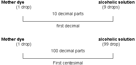
Acting like this, consecutively searing by the first
ones, we can get the subsequent decimal and centesimal potencies.Once we’ve performed such dilutions, the homeopathic
medicine must pass through a process called dinamization, that is, to such
the bottle tapping on its bottom with the palms (suction) or else, to
perform the same procedure in a special device assigned for this purpose.The term dilution must be reserved to the series of
successive operations described thoroughly in the several Organon
editions, and that allows decreasing more and more the quantity of
medicinal substances (Demarque, 1981).1.3. HOMEOPATHIC MEDICINE ORIGIN
The homeopathic medicine comes from the 3 reigns that
exist in the nature: animal, mineral and vegetal.As an example from the animal reign we have the snake
poison (Lachesis trigonogaster); from the mineral reign, the copper
(cuprum metallicum), the Gold (aurum metallicum) and the mercury
(mercurius solubillis), and finally from the vegetal, the belladonna
extracted from the plant Atropa belladonna. (Duprat, 1982).1.4. THE MEDICINE TESTED IN THE HEALTHY MAN
One of the Homeopathy’s principle is the
experimentation in the healthy man, what defines that the same medicine
that will Leal the sick one by the similarity Law (Tetau, 1980).This way, the practice of Homeopathy implies:
a) The experimentation of different substances
administrated in the healthy man. In this sense we can say that the
Homeopathy is an experimental method. The whole of the observed signals
during this experimentation, made obviously in sub toxic doses, gets the
name of pathogenesis. Some of pathogenesis comes from substances
unproved of toxic things (calcium carbonate, sea salt, silica). They
point out the notion of particular sensibility of certain individuals.b) Medicine in weak dose, and even very weak, rising
up many times beyond the number of Avogadro ( Tetau, 1980).Hahnnemann experimented several medicines, and after a
rigid observation, he catalogued the symptoms and signals produced by
these substances in his look Pure Medical Matter (Kent, 1980)1.5. THE ACTION OF THE HOMEOPATHIC MEDICINE
According to the Similatarity Law, the homeopathic
medicine, tested in the healthy man, causes characteristic physical and
mental symptoms, and they were registered by Hahnemann in his already
mentioned book Pure Medical Matter.It’s know that the choice of the homeopathic medicine
is made by a process of exclusion, unique and particular, in what the
patient’s symptoms must coincide as much as possible with the medicine.
That’s why it’s said that one is the mirror of the other (Tetau,
1980).The adage from Hippocrates similia similibus curantur
shows a precept that more clearly can be defined like this: the medical
substance able to establish in the healthy organism a set of analogous
disturbances that exist in the sick organism.The healing action of the homeopathic medicine is
revealed through a mechanism that can be analyzed according to two points
of view: pharmaco-dynamic, that reveals the duality of the action of all
medical substance according to the doses applied, and biological, that
interferes in the specificity of the organic defense (Duprat, 1982).The duality of action of all medicine has been known
for many years, and, not mentioning Heinnemann, we can find it in the
conclusions of several biologists, such as:Claude Bernard: “Every substance that, in small
doses, excites the qualities or functions of an anatomic element, in high
doses, repeals them”(Duprat, 1982).Brown-Séquard: “The moderate excitement of a
nervous element provokes exaltation of the functions that depend on it
directly or by reflex; a strong excitement can affect the same
functions” (Duprat, 1982).Hugo Schultz: “Every excitement provokes in a cell
increasing or decreasing of its physiological function, according to the
strong or weak intensity of the excitement” (Duprat, 1982).Huchard: “It’s enough to say that I held, long
time ago, only two therapeutic laws: the one from the treatment and cure
of a great number of morbid states with medicine that produce analogous
symptoms to the diseases, and the one from the prescribed medicine in
minimum doses, once it’s true, like Pecholier said, from Montpelier,
that in a single medicine there are several medicines, according to
different doses” (Duprat, 1982).Rudolf Arndt, according to the Fundamental Biology Law
states: ” The little increase it the strong ones subdue it, the
exaggerated ones, abolish it. This law is from the ” shaking
Law”, by Plunger, that fixes the action of the weak, medium and
strong electric current” (Duprat, 1982).Let’s appeal to some common examples, in order to
make concret these constant pharmaco-dymanics.“The opium, in strong doses, is a brain spinal
depressor and it produces a comatose sleep with muscular relaxation, in
small doses it stimulates the intellectual, nervous and muscular activity,
and it acts as a great excitant to keep person awaken” (Duprat, 1982)Talking about the arsenic, in its classical treaty of
therapeutics, Manquat says: ” The action of the arsenic in the real
gloves can be destructive, however, on the teeth is seems to act
differently. Actually in minimum doses, this medicine stimulates the
production of red globes, and in strong doses it’s destructive”
(Duprat, 1982).In short: “the effect of the strong does, which
Hehnnemann called primitive, and other authors called active, talks about
the action natural to the medical substance, its toxic virtue and a
coercive action, of denominated secondary or reactive, refers to action
natural to the organism, its defense reaction, its liberation from the
toxic threat of the medical substances” (Duprat, 1982).The IpecaCuãnha in strong doses is a vomitory, in weak
doses, diluted and strengthened, homeopathic, becomes one of the medicines
for nauseas and vomits. This way there is pharmacological inversion to a
certain dosage” (Tetau, 1980).1.6. HOMEOPHATIC MEDICINE EXPERIMENTATION
This research is based on the in vitro experimentation
of the following homeopathic medicines: Zincum metallicum, Cuprum
metallicum, Arsenicum Album and Manganum.1.6.1. ZINC
The zinc is a transition metal that performs a lot of
organic functions, being one of the main metals for the brain. It takes
part in the insulin formation as well as acts in the performance of almost
a hundred of enzymes of the system denominated zinc-finger, controlling
and activating a big quantity of hormones” (Halliwell and Gutteredge,
1989).The zinc is abundantly found in the prostate gland
secretion, in the seminal liquid, what suggests the cause and effect
relation between the zinc fault and the benign prostate gland
hyperplasia” (Handler, 1990).Plentiful in the nature, but combined with sulphur or
silica the Zincum metallicum can be found in France, England, India and
Australia.Even Hahnnemann who gave it us proper in therapeutics,
it has hardly ever been used in Medicine. Thanks to the homeopaths works
they could get good results from this remedy (Lathoud, 1980).1.6.2. ZINCUM METALLICUM
Zinc, used as a homeopathic medicine, gets the Latin
name Zincum metallicum.Zinc is a solid laminated metal, ductile, white bluish.
It’s fragile when it’s dry and heated until 200ºC, that’s why it’s
necessary to keep it at an intermediary temperature, when it’s exposed
to humidity it gets covered by a thin hydrocarbon cover, that adheres
strongly to the metal avoiding its oxidation. Heated until it’s live
red, burn to the air like beautiful green flame and comes to oxide of
zinc, Zn O. (Lathoud, 1980).Zinc acts on the set of sympathetic nervous system and
spinal brain. However its action falls back particularly on the thorax
plexus and on the big trunks and nervous branches that are distributed by
the locomotor system, that controls the movements and sensibility
(Lathoud, 1980).Zinc’s performance occurs in the motive part as well
as in the sensorial, above all in the nervous system. What iron is for
blood, zinc is for the nervous system ( Lathoud, 1980).1.6.3. COPPER
Copper is a transition metal that can be found in every
tissue, however in bigger quantities in the brain and liver.Copper acts as a co – factor in several enzymatic
processes, as catalytic converter in the synthesis of the hemoglobin, in
the conversion of tyrosine in melatonin of the thyroids hormones T3 and
T4, in the structural protection of the myelin sheath and in the elastin
and collagen synthesis. It also integrates the enzyme superoxide
desmutases, in the cytochrome oxidize in the tyrosinase and in the beta
hydroxilase dopamine (Torti, 1988).1.6.4. CUPRUM METALLICUM
Copper used as a homeopathic medicine gets the Latin
name Cuprum metallicum.Copper is metal with a characteristic red color, very
malleable, ductile and tenacious. At fresh air it’s covered by green
from hydrocarbon ate (grayish green). It’s found in the nature mainly in
the state of pyrite of copper and iron sulphate, often associated to
antimony, silver, lead and arsenic sulphates, and also in the state of
oxide and hydrocarbonate. It is also present in the most of vegetal and
animal food (Lathoud, 1980).Copper acts on the spinal medulla and the sympathetic
nervous system selectively, performing leading influence in the whole
body. It also acts, in the motor and sensitive enervations and in the
trophic enervation affecting the nutrition deep and directly ( Lathoud,
1980).1.6.5. ARSENIC
The arsenic is a toxic metal, whose contamination
sources are combustible oils, insecticides and tinctures. It’s
eliminated through urine.From the toxic metals, arsenic is one of the least
dangerous. However, the inorganic arsenics and the dangerous. However, the
inorganic arsenics and the trivalent forms are more toxic. Systemically,
when they are absorbed orally they can provoke vasodilatation in massive
doses, the arsenic can provoke deleteriores effects in the
cardio-circulatory system, like destruction of the capillaries and
arterioles, as well as myocardic necroses. In the gastro-intestinal
treatment it can provoke serious lesions like severe hemorrhages and
alterations of the cellular proliferation (Lathoud, 1980).The application in Homeopathy happened from the arsenic
toxicological observations on the human being, being used in these
circumstances, of course, dilested, when these effects are not observed.1.6.6. ARSENICUM ALBUM
The arsenic used as a homeopathic medicine gets the
Latin name Arsenicum album.The Arsenicum album, anhydrous arsenious, arsenic acid
or white arsenic, commonly called arsenic, whose formula is AS2O3, is the
most important from the arsenic compounds. It’s barely found in the
natural state, and generally it’s obtained by combustion of the iron
sulphurus arsenic or from other arsenifero minerals of cobalt or nickel.
It’s presented as a white powder crystallized, very similar to sugar,
inodous, slightly acid flavor, however a serious acridity is developed as
the time passes. Pulverized on the fire it decomposes and produces a
garlic smell. It’s soluble only in 82 parts of cold water, 140 parts of
alcohol to 95º C and 5 parts of glycerin. The 3 first pontentealizations
are generally obtained by trituration and the rest by, dilution (Lathoud,
1980).In general, the Arsenicum album is used as therapeutic
agent of great potency and diffusion. It has an immense field of action:
” it embraces the whole organism, and because of its elective
localization on the sympathetic nervous system it is meaningfully
affected. We can say that it irradiates to all the organic systems”.
(Espanet apud Lathoud, 1980).In a single word, the action of this unique
polychrestus is indefinite since the benign stages (weak irritation),
until the extreme cachexia (complete, chronic action). ( Lathoud, 1980).1.6.7. MANGANESE
The manganese is a transition metal, that takes part in
the molecule of mitochondrial superoxide dismutasis, from where one can
infer its relevant importance in the endogenous mechanism of stress
oxidative control and lipidic peroxidation (Torti, 1988).The manganese acts as a co-factor in the synthesis of
biotin, acetylcholine, cholesterol, thyroid hormones, thiamin, vitamin C
and prothorombin as well as acts as an activator of the peptidase, the
arginase and also in the glucose metabolism and in the absorption and
transportation of copper. (Hendler, 1990).1.6.8. MANGANUM
The manganese used as a homeopathic medicine gets the
Latin name Manganum.The manganum, white-grayish metal, hard, breakable,
inalterable when exposed to the environment, to normal temperature it is
presented under a petroleum cover. To hot air, it’s lightly revested by
an oxide cover. It disintegrates slowly in cold water, faster in
ebullition. Under pulverization this decomposition is very fast, even
under normal temperature. It can be found in many minerals in the state of
oxide silicate, phosphates, sulphurus, and sometimes in plants ashes, in
bones and also blood (Lathoud, 1980).In Homeopathy it is used pulverized metallic manganese
and with it the 3 first dinamizations are prepared by hahnemanneana
trituration (Lathoud, 1980).Manganese or Manganum aceticum, acts deeply on the
nervous system, the skin and the bones, followed by a serious anemia state
and intense debility.1.7. REACTIVE OXYGEN SPECIES – ROS
1.7.1 THE ORIGEN OF OXYGEN
The molecules of diatomic oxygen in the earth
atmosphere are the biggest ones to provoke reactions on the living cells.
Thus, it’s appropriate to start with some comments about, the oxygen,
only later we will have some considerations about the nature and
definition of free radicals.Except for those organisms that are specially adapted
to live under anaerobic conditions, all the animals and plants need oxygen
for an efficient production of energy (Halliwell and Gutteridge, 1989).The oxygen emergency must have been followed by the
ozone layer appearance (O3) in the high atmosphere, and the absorption of
the damage effects of the ultraviolet the evolution by the ozone, layer
probably allowed the evolution of the most complex earth organism
(Halliwell and Gutteridge, 1989).1.7.2 THE OXYGEN METHABOLISM
The oxygen is the element that, in the periodic
classification of the chemical elements, belongs to the family 6A, whose
atomic number and atomic mass are 8 and 16 respectively, and it possesses
8 electrons distributed in its orbitary layers.Normally, around 95 to 98% form the oxygen absorbed by
the aerobic organisms is reduced, forming water in the breathing chain
though the electrons transportation in the mitochondria as well as in the
endoplasmic reticulum, where the enzymatic system cytochome, in the
process of oxidative phosphoroclastic reaction proceeds the tetravalent
reduction of O2 by the cytochrome oxidase system, putting up
simultaneously 4 electrons to the oxygen, that it is directly reduced to
the water:O2 + 4H+
+ 4e– è
2H2O
The sources that give the cations of hydrogen and the
electrons to the reaction are, basically, the NADH, the FADH and the
ubiquinone or co-enzyme Q ( Halliwell and Gutteridge, 1989).However, as already referred, from 2 to 5% of O2
is reduced only 1 electron, and this one is going to occupy one of the
external orbitals, at the same time that the other keeps not matched,
producing intermediaries highly reactive, called Reactive Oxygen Especies,
that sometimes constitute the free radicals. This way, the first reactive
toxical specie of oxygen, the superoxide, like in the scheme:O2 + 4H+
+ 4e– è
2H2O1.7.3 FREE RADICAL
Free radical is any spice able of independent existance
and which contains one or more electrons unmatched, that is, electrons
present individualy in atomic or molecular orbitals (Halliwell and
Gutteridge, 1985).Na unmatched electron can join to isolated atoms
(hydrogen, metallic ions) or even to molecules (sugar, proteins, lipids,
DNA) what comes to be a process of biologic relevance (Slater, 1984 –
Halliwell, 1987).On the other hand the free radicals have already been
related to several human illnesses and they take part as fundamental
components in many of them, what shows us how big is the oxidative damage
caused by them (Halliwell and Gutteridge, 1985).1.7.4 HYDROGEN PEROXIDE
Another way of ROS is the hydrogen peroxide.
The hydrogen peroxide, although it’s not a specie
very reactive, it is na agent able to inactivate enzymes, mainly by
oxidation of essential thiol groups. Its bigger oxidative power, however,
is indirect, as the generator of the hydroxil radical (HO·
) and by the interaction with the superoxide radical (O2·
–). In this case it’s a powerful oxidative, and in
enough concentrations, it can kill can cells, and in the presence of iron
its toxicity can increase from 10 to 100 times (Eaton, 1991).There are 2 kinds of enzymes that remove the hydrogen
peroxide. They are the catalase and the peroxidase glutathione ( Halliwell
& Gutteridge, 1989).Under the peroxidase glutathione action, the hydrogen
peroxide reacts with the reduced glutathione, oxidate, and forms 2
molecules of water and oxidated glutathione.GSH – Px (selenium)
2 GSH + H2O2 <---------->
GS-SG + 2 H2OGlutathione redutase
The oxidated glutathione is once again reduced
(regenerated) by the action of the redutase glutathione enzyme. We can
notice that for the perfect working of the peroxidase glutathione, the
presence of sellenium is fundamental, and for the redutase glutathione
action there must be vitamin B2.Under the catalase action, there is also the formation
of water, besides the molecular oxygen, So we have the following reaction:Catalase
2 H2O2 ———-> 2 H2O2 +
O2The HO can be generated in the cells by exposition to
the ionizant radiations, from other species of oxygen, as well as by the
Fenton and Haber-Weiss Reactions, mediated by metallic ions.1.7.5 FENTON REACTION
The mixture of hydrogen peroxide and Fe2+ reacts to
many organic molecules, as was firstly observed by Fentom, in 1984. The
reactivity is due to the hydroxil radical formation (Halliwell and
Gutteridge, 1989).Intermediary complex
2 H2O2 + Fe2+
———-> OH– + OH·
+ Fe3+
Traits of Fe3+ are available to react again
with the H2O2, although such a reaction is very slow
in physiological pH:Intermediary complex
Fe3+ + H2O2
———-> Fe2+ + O2–
+ 2H+
The simple mixture of the with the hydrogen peroxide
can ceirtainly form, in the biological system, under some conditions, a
range of oxidative reactions (Halliwell and Gutteridge, 1989).1.7.6. HABER-WEISS REACTION
This reaction occurs in the presence of iron or copper
salts, like the following:O2· – +
Fe3 ———-> O2 +
Fe2=> O2–
Iron salt
Fe2+ + H2O2
———-> Fe2 + OH– +
OH·Or copper
The ferrous complexes are able of the hydroxil radical
formation (OH), as they present a low molecular mass, besides the O2·
– and the H2O2 facility of
liberating iron ions active in a catalytic way, coming from proteins. Thus
the increase of the O2· –
and the H2O2 generation can creat conditions to the
formation of the OH. (Halliwell and Gutteridge, 1985).1.8. OTHER PHYSIOLOGICAL CONDITIONS
The reactive species can also be produced by the
utilization of the diet compounds (Ames, 1989).Following we have the table with the main reactive
oxygen species (Sies, 1991).TABLE 1: Reactive Oxygen Species – ROS
O2· –
Superoxideânion ou superoxide radical
HO2·
Perhidroxil radical
H2O2
Hydrogen peroxide
OH·
Hidroxil radical
RO·
Alcoxil radical
ROO·
Peroxil radical
ROOH
Organic hidroperoxide (e.g.: lipoperoxide)
1
O2
Singlet oxygen
RO·
Excited carbonil
1.9. ROS AND ANTIOXIDANT SYSTEMS
Not all the ROS have systems that make them inactive.
For some ROS there are endogenous desativator systems, while for the
others there are external antioxidants, or even both.The external antioxidants are also called scanvengers
or ROS sweepers (Halliwell and Gutteridge, 1989).Following there is the ROS table related to their
correspondent antioxidants.TABLE 2: Reactive Oxygen Species and Antioxidants
ROS
ANTIOXIDANTS
ENDOGENORES
EXOGENORES
Superoxide (O2· –)
Superoxide Dismutasis:
a)Cytophasmic: zinc-Copper
b)Mitochondrial: Manganese
Vitamins, zinc, copper, manganese, pycnogenol, ethylene diamine
tetra-acetic acid
Hydrogen Peroxide (H2O2)
Catalase Fe2+
__________
Lipidic Peroxide (COOH–)
Glutathione Peroxidose, selenium
Vitamin E, selenium
Hydroxyl Radical
(HO·
)
__________
Vitamin C, pycnogenol, dimethil sulfoxide, ethylene
diamine-tetra-acetic acid, dimercapto succinic acid and manitol
Singlet Oxygen (1O2)
__________
Beta carotene
1.10. ROS FORMATION
The ROS can by very toxic when excessive, either by
elevated production or by organism difficulty in neutralizing them. They
are formed starting by the oxigen, what is a paradox, because at the same
time that it generates and support the life, on the other hand it can be
lethal (Halliwell and Gutteridge, 1989).Firstly the superoxide radical (O2·
–) is formed, which can be dismuted in hydrogen peroxide
(H2O2) or even through catalytic action, by the
acting, of the superoxide dismutasis enzyme (SOD).In the organism there are two main SODs, one of them
cytoplasmic, that is the CuZnSOD, and the other mitochondrial, that is the
MnSOD, this on containing manganese and that one containing Copper-Zinc in
the same molecule.The importance of the SOD can be shown by the fact of
being the most abundant enzyme of the organism, as well as it also the
fifth most abundant protein in the same organism. (Halliwell and
Gutteridge, 1989).The following graphic shows how the ROS formation
occurs.

SOD – Superoxide dismutasis, Cat – Catalase, O2·
– – Superoxide, O2 – Oxygen
GSSH – Oxidate Glutathione, GSHPx – Glutathione Peroxidase, H2O
– Água,
OH· – Hydroxyl Radical, GSH – Reduced
Glutathione, H2O2 – Hydrogen Peroxide.
1.11. ROS ACTIONS IN THE BIOLOGICAL SYSTEMS
The man is probably the result of evolutionary
processes of unicellular and anaerobic organisms. The terrestrial
environment gained more complex organisms only layer development, which
allowed the absorption of part ultraviolet solar radiation, limitative
factor for life, that for a long time, was confined to the aquatic
enviroment. Whith the ultraviolet radiation reduction, which causes
damages to the living beings, the terrestrial environment became
compatible to life, unchairing the acceleration of the evolutionary
process (Halliwell and Gutteridge, 1989).As a result of these changes, the man remained with
remanescent of his anaerobic system, that’s why the oxygen one breaths
– and that is so important to maintain the life in different
circumstances – can be the cause of death or diseases development, as it
is one of the most important generators of free radicals inside the
organism (Halliwell and Gutteridge, 1989).The ROS, as they react to the majority of the organism
molecules, they are able to interfere in the biological processes, causing
several diseases, mutations, oldness, among other alterations (Halliwell
and Gutteridge, 1985).1.11.1. ISCHEMIA AND REPERFUSION
The ischemia comes from the initial obstrution of the
blood flux, for exemple, due to the arterial clamping for a certain period
of time, what provokes tissue suffering by hypo-oxigenation. With the
unclamping, the tissue reperfusion occurs, determining the brusque arreval
of oxygen and nutriens to the tissue, with a meaninful encrease of free
radicals, extreme important phenomenon, mainly during the first 60 to 90
seconds, because it damages the reperfused area, provoking micro-heart
attacks (Haliwell and Gutteridge, 1989).Tissues that suffered hypoxia or ischemia survive for
variable periods, depending on the nature of the tissue involved.It the period of ischemia or hypoxia is short, the
tissue damage can be minimized with the re-introduction of nutrients. The
re-introduction of O2 in the ischemia or long tissue hypoxia –
can cause – aditional tissue injury (re-oxygenation injury) which is
partiatly mediated by oxygen radicals (Halliwell and Gutteridge, 1989).The ischemia and reperfusion may occur in the cardiac
surgeries, where the re-oxigenation can cause serious arrhythmias with
extense lesions due to the oxygen paradoxal shock, which causes ATP
depletion during the ischemia, forming hypoxanthine or xanthine, that work
like a substrate to the xanthine oxidase enzyme (XO), precursor form the
uric acid, that acts like na antioxidant in these cases.During the re-oxigenation, through the reperfusion, the
free radicals production occur with its deleterious effects, what makes
the ischemia even worse (Halliwell and Gutteridge, 1985).The xanthine oxidase is produced from the xanthine
dehidrogenase in the presence of the ion Ca2+, generating free
radicals that will damage the tessues, that’s why the use of
antioxidants like the SOD or the manitol is commended before the arterial
inclamping.1.11.2. NEUROLOGICAL DISEASES
It has been observed that cranium-encephalic
traumatisms cause cerebral injury or spinal medulla. The lead can provoke
degeneration that involves reactions with free radicals, like the
lipoperoxidation.Cerebral ischemia or hypoxia, followed be reperfusion,
must also estimulate the lipoperoxidation (Halliwell and Gutteridge,
1989).The brain contans 3 important characteristics:
1. It is very rich in polyunsaturated fatty acids
that act like substrate to the lipidic peroxide formation;2. It is free iron richest organ, which acts forming
the radical hydroxyl;3. It’s formed by cells that do not reproduce
themselves, the neurons, that reach the maturity around 30 years old
and, later, they will be lost at about 10.000 to 100.000 a day.Another mechanism that contributes to the cerebral
injury after the hypoxia ischemia is the non production of exciting
neurotransmitters animoacids, like the glutamate or aspartate ( Halliwell
and Gutteridge, 1985).In the Parkinson disease, most part of the works has
shown there are free radicals increase, mainly in the brain’s nucleus
that will produce L – dopa and are mainly cytotoxins and neurotoxins,
that cat at this level increasing the superoxide production (Halliwell and
Guteridge. 1985).The Alzheimer disease, senile insanity, presents some
proper characteristics, like: neurofibrillar tangles, ghost neurons,
lipoferous deposit (pigment formed by the malondialdehyde connected to the
protein), matched filaments, amyloidosis (the amyloidosis protein
precursor, APP, is a potent free radicals generator), increase in the
activity of the neuritic plaques and oldness of some parts of the brain
like: raphe neclei, hyppocampus and locus ceruleus (Halliwell and
Gutteridge, 1985).The aluminium is the most abudant metal of the
terrestrial crust and we are constantly exposed to it. The presence of
high aluminium concentrations in the brain of patients with alzheimer
suggests this is one of the causes of the causes of encephalopathy present
in the disease (Halliwell and Gutteridge, 1985).Two of the pathological characteristics of the
Alzheimer dementia, firstly observed by Alois alzheimer in 1906, are the
presence of senile plaques and neurofibrillar tangle in the brain. The
aluminium action must contribute to the neurotoxic proprieties, as we know
the brain is sensitive to the free radicals reactions (Halliwell and
Gutteridge, 1989).1.11.3. LIPIDIC PEROXIDATION
The lipidic peroxidation is the process through which
the ROS attack the polyunsaturated fatty acids of the membranes
phospholipids of the cells, desintegrating them and allowing, this way,
these species entrance in the intracellular structures.The phospholipases, activated by the toxic species,
desintegrates the phopholipids, liberating the non saturated fatty acids
(Halliwell and Gutteridge, 1989, resulting in the following deleterious
actions of the lipidic peroxides:
- cellular membranes rupture (NA/k and Ca/Mg bombs)
- DNA mutations – deoxyribonuclei acid
- Unsaturated lipids oxidation
- Chemical residues formation like the malondialdehyde
- Components engagment of the extracellular matrix, proteoglycans,
collagen and elastinThe lipidic peroxides possess na action power higher
than the other primary toxic species of O2 (O2·
–, H2O2, OH·
, O2), reaching further targets easily.The lipoperoxidation must also have a very important
role in the cellular proliferation, especially in tumoral cells. Some
authors suggest that the lipoperoxidation products are involved in the
cellular division control. On the other hand the lipid peroxidation is
related to the tumoral increase (Gonzalez, 1992).1.12. ANTIOXIDANT MECHANISMS
The oxygen is a paradox in the planet, because it is
essential to live as well as it can cause injures to the organism
(Halliwell and Gutteridge, 1985).The antioxidant agents can’t distinguish between the
reactive oxygen species that have a physiological role and those that are
causing damage. Because of this, their action can, in some not be
profitable to the organism. However it is with the balance between the
oxidant and antioxidant species that the organism, will be able to obtain
the conditions to a better performance of its functions, considering that
a disturbance in this balance can result in a range of pathological
processes (Bast et al, 1991).In general the following strategies have to be followed
with the intention of increasing the efficiency of the OA.Oxidative Stress
=====>> Metal Complexes descompartimentalization
=====>> Excessive production Of O2·
– (superoxide)
=====>>Anti-Radical defenses reduced1.13. ANTIOXIDANT DEFENSE
The antioxidants that represent the organisms defense
against the reactive oxygen species are divided in two main kinds, the non
enzymatic and the enzymatic.1.14. NON-ENZYMATIC ANTIOXIDANTS
Some essential nutrients can attack directly the oxygen
radicals. The vitamin E (alfa-tocopherol) is the biggest liposoluble
anti-oxidant present in all the cellular membranes, and therefore, acts in
the protection against the lipoperoxidation (Kay et al., 1986). It can
react directly with a variety of oxiradicals, like the superoxide, the
hydroxil, etc, and also with the singlet oxygen ( Machlin and Bendich,
1987).The vitamin E was firsty related in 1922, in the USA,
by Evaris and Bishop, who demonstrated it is liposoluble and also
essential factor for the normal reproduction in mice. The purification of
this factor revealed it is compound from the tocopherol family. Four
tocopherols are known, yet the alfa-tocopherol is the most improtant
biologically and the terms alfa-tocopherol and vitamin E (Halliwell and
Gutteridge, 1989).First of all the inactive alfa-tocopherol reacts with
the singlet and could, this way, protect the membrane against this
specie.(Halliwell and Gutteridge, 1989).During its antioxidant action like chain – breaking
(destroying the lipoperoxidation chain) in the membranes the
alfa-tocopherol is consumed and converted in form of radical (Halliwell
and Gutteridge, 1989).The vitamin E can also protect against the
peroxidation, modifying the membrans structure (Halliwell and Gutteridge,
1989).The vitamin E, situated near the cytocrome P-450 in the
membrane phospholipid, sweeps the free radicals formed in the cytocrome
P-450. Then, the vitamin C reduces the radical tocopheril. In a recent
study held at Tufts University, it was proved the powerful imuno-stimulant
action of the vitamin C. (Halliwell and Gutteridge, 1989).The vitamin E, called the vision vitamin was discovered
at about 2.000 years, when the Greeks verified that the animals’liver
had something that cured some eyes affections, that’s why the name
retinol, due to its importance for the vision. It was the first vitamin to
be cataloqued, therefore the name A. (Halliwell and Gutteridge, 1989).The retinol is essential in the human diet and it is
usually known as vitamin A, one of the liposoluble vitamins (Halliwell and
Gutteridge, 1989).The carotenoids, mainly the beta-carotene can work as
vitamin A precursors. They are absorved by the human bowels and must also
actuate like antioxidants. They have a double role, they decrease the
singlet oxygen formation in vivo, and help to remove those already formed.
(Halliwell and Gutteridge, 1989).The vitamin A has little antioxidant action and it is
unable to act on the singlet oxygen, but its precursor, the beta carotene,
is the most efficient linking of this reactive form of oxygen found in the
nature and it can act like antioxidant, the beta carotene, a pigment
present in all plants, can be found in cellular membranes, including in
the lipossomes (Machlin and Bendch, 1987).The pure ascorbic aced is soled, white, crystalline and
very soluble in water. Plants and animals can synthesize it, except the
humans, primates and cavies that can’t synthesze it and need to obtain
it from the diet (Halliwell and Gutteridge, 1989).The ascorbic acid is necessary in vivo like cofactor of
several enzymes, and the most well-known are the proline hydroxylase and
the lysine hydroxylase, involved in the collagen bio synthesis. The
ascorbate deficiency in the human diet causes the scurvy . The most
impressive chemical propriety of the ascorbate is its alility to act like
reducer agent (electrons donor). (Halliwell and Gutteridge, 1989).The vitamin C (ascorbic acid), is hydrosoluble and also
acts against the free radicals and the singlet oxygen. The ascorbic acid
also takes part of the regeneration of the vitamin E antioxidant and
reduced form (Halliwell and Gutteridge, 1985).The vitamins A, C and E, can also be found in high
concentrations in the plasma, in the adrenal glands, in the brain, liver,
and in the blood cells among other regions (Porta, 1988).The is also a range of other non-enzymatic antioxidants
that take part in the defense against the reactive oxygen species in the
biological systems like, for exemple, the ubiquinone, the ceruloplasmin,
the uric acid, the taurine, the flavonoids and other fenolic compounds of
vegetal origin (Halliwell 1990, Cutler, 1991, Sies, 1991).The ubiquimone is used a lot to improve the cardiac
function in the cases of congestive cardiac insufficiency, myocardium
ischemia, angina pectoris and arterial hypertension. It has been used in
the multiple sclerose and the Alzheimer diease. It also acts in the
mellitus diabetes, the periodontic diseases and muscular dystrophies, as
well as in the imunological system disfunctions (Halliwell and Gutteridge,
1985).The glutathione (GSH) is a cellular health marker and
its fall indicates oxidant lesion. Its deficit causes decreasing in the
resistance to drugs and radiations, the capacity of tumor reversion and
the ascorbate syntheses in animals (Halliwell and Gutteridge, 1985).The glutathione is a tripeptide composed by
non-essential aminoacids, describe as an important antioxidant agent
(Ames, 1983, Halliwell and Gutteridge, 1985).As we could infer from the chemistry of the oxygen, the
univalent reaction, generates O2.-. The main escape system is observed
thorgh the complex NADH- Coenzyme Q redutase and from the reduced forms of
the coenzyme Q10. (Halliwell and Gutteridge, 1989).The human neutrophil and several other tissues (nervous
tissue, for example) are taurine-rich. There are several propositions
saying the taurine has a biological role and acts as an antioxidant
(Halliwell and Gutteridge, 1989).1.14.1. THE MELATONIN
The melatonin is a well-known antioxidant, produced by
the pineal and gland and its main hormone was discovered by herner in
1958. It is also produced in other tissues, like retinal and large
intestine. (Guyton, 1973).The pineal gland is the first endocrine gland that is
formed in the embrional phase. Its works like a “biological clock,
when it secretes the melatonin at night, united to other neuropeptides
(Maffei, 1978).The melatonin secretion is ten times bigger at night
than during the day. This concentration reduces as the time passes,
reaching the top in the adolescence. In the elderly it corresponds to half
comparing to the yoyngsters (Smith and Tier, 1990).Chemically, the melatonin is nominated by 5 acetyl –
5- methoxy-serotonin and it is derivative from the essential aminoacid
tryptophan, found in proteinous food like grains, seeds and vegetables.Recently, the melatonin was described as part of the
immine functions of the organisms and as a mighty antiioxidant. Performing
the antioxidant function, the melatonin seems to unchain a substantial
protection against the free radicals which are generated in a variety of
experimental situations, including the lesion for ischemia reperfusion.
That’s why it has been used therapeutically in surgeries and transplants
(Reiter and Maaestroni, 1999).1.14.2. MINERALS
Beside these, there are several essential nutrients
from mineral origin, that take part in the antioxidant process in
association with enzymes. They are: zinc, copper, manganese, selenium and
iron (Halliwell and Gutteridge, 1985).1.14.3. ENZYMATIC ANTIOXIDANTS
Antioxidants are any substances that when they are
present in minor concentrations, compared to those oxidizable substrata,
meaninfully delay or inhibit this substratum oxidation and they can act in
different levels of the oxidative sequence (Halliwell and Gutteridge).In 1954, Gershaman and Gilbert proposed that most of
the harmful effects caused by the high oxygen concentrations in live
organisms could be attributed to the free radicals formation. However this
idea didn’t raise many researcher’s interest until 1968 with the
discovery of na enzyme that is specific for the catalytic removal of na
oxygen radical (MCCord and Fridovich, 1969). This enzyme called superoxide
dismutases, along with catalase and glutathione peroxidase are the main
antioxidant defenses that act in the superior organisms (Halliwell and
Gutteridge, 1989).1.14.4. ENZYMES
The enzymes: superoxide dismutase, catalase and
glutathione peroxidase represent the main endogenous defense of the
organism.1.14.4.1. SUPEROXIDE DISMUTASE
It’s possible that the superoxide dismutase (SOD) be
a substance with real anti-aging effects, and it can act positively over
all the degenerative processes (Hendler, 1990).The superoxide dismutase (SOD) has a fundamental role
in the organism defense against the reactive oxygen species because it
acts removing the superoxide. Before it was discovered, the SOD had
already been described by some authors as a protein that contains copper,
but they hadn’t attributed to it any catalytic activity (Halliwell and
Gutteridge, 1985).After McCord work however (1969), with the
determination of its function in the superoxide radical dismutation (O2·
–), its role was established and even today, although
there are many researches with this enzyme, no other substrate was
described, showing its specificity to the superoxide (Halliwell and
Gutteridge, 1985).In 1938, T. Monn and D. Keilin in England, described a
blue-green protein, isolated from the bovine flood that also contained
copper and called it hemocuprein. In 1953, a similar protein was isolated
from the horse’s liver and called hepathocuprein. Other proteins of this
type were isolated, like the brain – cupreine from the brain. In 1970,
it was discovered that the proteins from the erytrocyte contain zinc as
well as copper. No enzymatic function was detected in any of these
proteins, so it was suggested that they served like metals deposit.
However, in 1969, Jim Cord and J.Fridovich’s work, in the USA, showed
that the protein from the evythrocyte can remove in a catalytic way the
superoxide radical, and thus this function was identifed as the superoxide
dismutase enzyme (Halliwell and Gutteridge, 1985).There are different kinds of SOD, depending on the
metal that acts like a co-factor in its catalytic site, but all of them
act basically according to the same reaction described by McCord and
Fridovich in 1969:
O2· – +
O2· – +
2H+ è O2 +
H2O21.14.4.1.1 SUPEROXIDE DISMUTASE COPPER-ZINC DEPENDENT
The form that contains copper and zinc, called
superoxide dismutase copper-zinc dependent (CuZnSOD), is very stable and
seems to be present in practically all the eukaryotic cells (plants or
animals). (Halliwell and Guttiridge, 1985).On the other hand, in prokaryotic cells like seaweeds
and bacteria, the catalytic activity related to CuZnSOD seems to be
restricted to the symbiosis of these organisms with eukaryotes where the
enzyme is present (Fridovich, 1978. Halliwell and Gutteridge, 1985).The eukaryotic CuZnSOD also called SOD A, has a
molecular weight of 32000 and it is formed by two identical proteinous
sub-unities, with on atom of copper and one of zinc in each one. It’s
the cytoplasmic form of the SOD, and its properties have been really
resisting to the evolutive changes, beingle able to distinguish the enzyme
gotten from fungi, plants, fowls and mammals easily (Fredovich, 1977).The copper takes part in the dismutation reaction
passing alternately by oxidation and reduction, like the exemple:
Enzyme – Cu2+ +
O2· – è
E – Cu + O2E – Cu +
O2– + 2H+ è
E – Cu2+ +
H2O2O2– +
O2 + 2H+ è
H2O2 + O2
(resultante reaction)At last, another action mechanism is also possible
in what the first O2– does not reduce the copper
ion, but forms a complex with it.The Zn does not work in the catalytic site, but is
appears to stabilize the enzyme. This conclusion was obtained from
experiences in which the metals were removed from the active sites and
replaced in others, alone or in groups (Halliwell and Gutteridge, 1985).1.14.4.1.2 SUPEROXIDE DISMUTASE DEPENDENT ON THE
MANGANESE (MnSOD).The superoxide dismutase dependent the manganese
(MnSOD) is a pink protein, whose molecular weigh is 40.000 and it contains
manganese in the active sites. Its activity decreases in alkaline pH. It
is not inhibited by the cyanide neither by di-etil-di-hydrocarbonate. It
is destroyed by the chloroform + ethanol (it does not survive to the
typical methods of the purification to the CuZnSOD). The MnSOD activity
related to the CuZnSOD depends on the tissue and species vhere they act.
The Mn removal form the active sites causes catalytic activity loss, and
it cannot be replaced by any transition ion, because it loses its
functional activity. The aminoacid sequences of all the MnSOD, in all the
species, are allke, and they are not related to the CuZnSOD (Halliwell and
Gutteridge, 1989).The superoxide dismutese manganese dependent (MnSOD),
found in bacterial like E-coli and Streptococus mutans (Mc Cord et al.,
1971, Fridovich, 1978), does not seem to have any relation with the
CuZnSOD included in the eukaryotes cytoplasm, except by its catalytic
activity.There is also the mitochondrial superoxide dismutase or
SOD B in human tissues (Beckman et al., 1973), that is similar to the
prokaryotes MnSOD but it has four sub-unities instead of two, and it has a
molecular weight of about 80.000, having also a manganese atom per
sub-unit. The mitochondrial form of the SOD is much more similar to the
prokaryotes MnSOD than to the CuZnSOD (Fridovich, 1978).1.14.4.1.3 EXTRA CELLULAR SUPEROXIDE DISMUTASE
Finally, there is one more form of SOD in human tissues
different from the others already described. This enzyme has molecular
weight of 135.000 and it is composed of four iqual sub-unities more
covalently connected. Each molecule seems to contain four copper atoms,
and iron or manganese are not found in the enzyme. Because it is present
mainly in extracellular fluids like the plasma it was nominated
estracellular SOD (ECSID). Its activity is not very significant compared
to the other superoxide dismutase forms (Markeund, 1982).1.14.4.1.4. PARTICULARITIES
Related to the catalytic activity of the SOD different
forms, we can assure the CuZnSOD is reversibly inhibited by the cyanide
and by H2O2 in concentrations over 10 uM (Fridovich,
1978). It can also suffer inactivation by the exposition to the superoxide
radical, while the MnSOD is not inactivated in the same conditions (Senet
et al, 1981).The CuZnSOD form is more resisting to temperature
variations and to denaturing by substances like guanidine chloride,
dedecyl sodium sulphate, or urea (Halliwell and Gutteridge, 1985). It’s
also the most resisting to pH variantions (Fredovich, 1978).The immunological tests have been more used to the
protein quantificatton or CuZnSOD and MnSOD. As these proteins are very
different, the antibodies do not make crossed reactions. The tests are
limited in human tissues to they serve as master lines. They are better
evaluated in the liver, because there is a bigger concentration of SOD
(Halliwell and Gutteridge, 1989).1.14.4.2. GLUTATHIONE PEROXIDASE
The glutathione peroxidase enzyme (GPX) was discovered
by Mills in 1959, in mammals’tissues. It is not present in plants or
bacteria, although it can be found in some seaweeds and fungi (Halliwell
and Gutteridge, 1985).The substrate to the GPx is the tripeptide glutathione,
found in the most of animals, plants and even in some bacteria. The enzyme
catalyses the reduced glutathione oxidation (GSH) to oxidanted glutathione
(GSSG) using the hydrogen peroxide:H2O2 +
2GSH è GSSG + 2H2OThe maintenance of GSH levels occurs by the action of
the glutathione redutase enzyme (GSSG), once again in its reduced form
(Cohen and Hochstain, 1963; Paglia and Valentine, 1967):NADPH ———-> GSSG +
H+ è 2GSH ———-> NADPAlthough the GPx is specific for glutathione as
hydrogen donor, it can accept other peroxides besides the H2O2
(Beutler et al, 1974; Halliwell adn Gutteridge, 1985), in reaction
that can be shown like:2GSH + R-OO-H è
GSSH + R-OH + H2OThe animal cells have two kinds of glutathione
peroxidase, and one of them is selenium dependent while the other is not.The first kind is able to reduce any organic
hydroperoxide , besides H2O2. This form has a
molecular weight of 81.000, it is a tetrameric protein and it has one atom
of selenium in each sub-unit (Forstron et al, 1978; Meera Khan et al,
1984; c Bridge et al, 1988).The relation between the GPx activity and the selenium
supply in vivo has been object of lots of clinical and experimental
investigation. In human beings, the enzyme genic regulation in lineage of
myeloid cells (HL-60) showed to happen pos-transcriptionally being
controlled by the selenium availabelity (Chada et al, 1989).The second kind, which is not selenium dependent, has a
molecular weight of 35.000, it is dimeric and it is able to reduce any
organic hydroperoxide, except the H2O2 (Meera Khan
et al, 1984; Ciriolo et al, 1991).The GPx has high activity in the liver and
erythrocites, while in the brain, heart and lungs its activity is
moderate, and in the muscle it is very reduced (Cohen and Hochstein,
1963).The GPx finds high activity in the liver, moderate
activity in the heart, lungs and brain, and low activity in the muscles
(Halliwell and Gutteridge, 1989).In the most of animals, the selenium dependent enzyme
is responsible for the major part of the GPx varies a lot among the
different species as well as from tissue in the same specie (Mannervik,
1985).In mice, the GPx distribution has been widely studied
and, in hepatocites the GPx selenium dependent is located mainly in the
cytosol and in the metochondrial matrix (Mannervik, 1985),The good pH for the GPx is next to 8.0 but the enzyme
keeps active with high values. Its activity is minimum in pH lower than
6.0 (Mills, 1959; Paglia and Valentine, 1967).1.14.4.3. CATALASE
It is an enzyme that is present in most of the aerobic
cells, and in animals it is found chiefly in the liver, kidneys and
erythocytes. However organs like brain, heart and skeleton muscle contain
small quantities of the enzyme (Halliwell and Gutteridge, 1985; Masters
and Crane, 1990).In mammals, like rats and mice, the most part of the
enzyme catalytic activity happens in the peroxisomes, and only a small
part has a cytoplasmic or reticular origin (Master and Crane, 1990; Percy
1984).In 1818, Thénard had already observed that the
hydrogen peroxide was decomposed by animal tissues, with oxygen discharge.
After 83 years, Loew established that this effect was due to a specific
enzyme called catalase. Warbing in 1923, suggested that the catalase has
iron atoms. In 1990, Geili and Helestrom found evidence that its prostetic
group was the humatin (Percy, 1984).The catalase mechanism of action can be synthesized
this way, according to chance ( 1952 a and b):Catalase – Fe (III) + H2O2
è compound ICoumpound I + H2O2
è catalase – Fe
(III) + 2H2O +
O2If it is not neutralized, the H2O2
interacts with iron cations (or copper), originating the ion hydroxil
(inert) and the free radical hydroxil (active).Although the knowledge about the catalase mechanism of
action in vitro is old, until 1971 the information about its biological
role in vivo was still considered undevoloped (Chance and Oshino, 1971).The most important nutrients that are coadjutant are
the iron and the tocopherols (vitamin E), which are distributed in the
cellular membraine, in the hydrophobic phase (Halliwell and Gutteridge,
1985).After the identification of the primary intermediary of
associated enzymes with the hydrogen peroxide in the fractions of rat’s
liver peroxisomes, rich in mitochondrias, it was possible to establish the
function of this enzyme-substrate complex also in the biological systems,
considering that this action is considerably peroxidative (Chance and
Oshino, 1971).The catalase avoids the metahemoglobin accumulation and
it decomposes the hydrogen peroxide, a toxic product from the metabolism,
in water and molecular oxygen (Chance and Oshino, 1971; Wieacker et al,
1980; Gaetani et al, 1989).Beside its role as reactive oxygen specie and
therefore, causer of oxidative stress, the H2O2
surplus causes the hemoglobin oxidation and, thus, diminution of the
oxygen concentrations, what can cause infections, ulcer and even necrosis
(Wieacker et al, 1980).Despite the reactions involving the catalase have been
studied since the last century, the exact mechanism of action of the
enzyme still causes argument and its biological role keeps being
researcheg. It is peroxide elimination, the catalase also acts in the
oxidation of electrons donors, like ethanol, methanol, phenols, DOPA,
epinephrine, when the H2O2 has low concentrations
(Percy, 1984).The catalase enzyme has molecular weight of 240.000,
and when it is purified, it presents 4 subunits, each of them with a
grouping (Fe III- protoporfirin) connected to its active site (Keilen and
Hartree, 1945; Wieacker et al, 1980).The molecule dissociation in its sub-units causes loss
of its catalytic activity. It happens easily when the enzymes is stored
with freezing or infreezing, or by exposition to acids or bases (Keilen
and Hartree, 1945; Chance, 1952 a; Chance, 1952b; Percy, 1984).The catalytic activity of this enzyme can be, inhibited
by superoxide, azide, hydrogen cyanide (HCN), but it is not inhibited by
other cyanide ions (CNT) (Chance, 1952b). The most used inhibitor,
however, it is the triazol amine that acts over the compound I, when the
presence of the hydrogen peroxide is necessary so that the inhibition
happens (Halliwell and Gutteridge, 1985).1.14.4.3.1. INTERACTION
Recently, it was described the catalase inhibition by
the glutathione and by other compounds with thiol grouping, like the
dethiotreitol (DTT), and the reduced forms of these compounds have a
bigger unhibitory action than the oxidative (Sun and Oberley, 1989).Relating to the pH we can observe a reduction of the
enzyme activity under Ph 4.0. In the level of 4.0 to 8.5 the catalase
activity remains constant, and above this level it reduces again (Chance,
1952).Experiments have been carried out to evaluate the
competition between the catalase and gluthathione peroxidase enzymes in
erythrocytes.According to some authors the catalase is the enzyme
that makes the conversion of high H2O2
concentrations into water and oxygen; when the hydrogen peroxide is
present in low concentrations (normal physiological conditions), however,
the glutathione peroxidase is in charge of transforming it into water
(Cohen and Hockstein, 1963, Sinet at al, 1975; Halliwell and Gutteridge,
1985).Gaetani et al (1989) assured that the catalase and the
GPx perform the same activity in human erythrocytes, and that the
decreasing of one of them wouldn’t cause deleterious effects.Trying to establish the importance of the erythocytary
catalase, Scott et al (1991) used normal and acatalasemic human cells to
verify if in fact, the enzyme has a secondary role in the hydrogen
peroxide metabolism, like several authors assured (Cohen and Hochstein,
1963; Sinet et al, 1975, Halliwell and Gutteridge, 1985). According to
them, the use of acatalasemic cells proved the great importance of the
catalase which can be the first in defense of the H2O2,
once the GSH increasing or reduction (which would allow the elevation of
the GPx activity) didn’t alter the general antioxidant activity in the
normal or acatalasmic cells.Both systems seem to have advantages and disadvantages
to the organism while the GPx is more efficient (it has more affinity by
the substrate), multi-functional (it reduces free H2O2
and also other peroxides), slow (limited by the GSH recycling) and
metabolically expensive, the CAT has low affinity by the substrate but it
is extremely fast (Eaton, 1991). Thus the CAT must protect the calls of
large H2O2 quantities and the low endogenous levels
must be in charge of GPx, along, with the enzymatic system GSH dependent
(Eaton, 1991).1.15. OLIGOELEMENTS
Forsen, in 1897, elaborated the first scientif
definition of oligoelements: “oligoelements are chemical elements
which are present in the living matter in concentrations equal or inferior
to 0,01% of the dry corporal weight of the human organism”(Torti,
1988).Forsen’s definition is undoubtedly very important,
fowever only from the quantitative point of view, because it doesn’t
refer to the metabolic and biochemical processes of the oligoelements
(Torti, 1988).1.15.1. QUANTITATIVE DIFFERENCE
Ahead Forsen’s definition, it was verified the
existence of the essential oligoelements, that is, indispensable to life.To be considered as essential, an oligoelement must
have the following characteristics:1. it must be present in all healthy tissue of all
living organism;2. its tissue concentration must be relatively
constant;3. its lack induces to physiological and structural
alterations of many kends;4. it prevents or corrects the morbid affections,
caused by its lack ( Torti, 1988).It is interesting to emphasize the similarity or
analogy between the oligoelement and the vitamins, mainly concerning to
itens 1 and 2, because the lack of vitamins also induces to functional and
structural alterations, and they can be prevented or corrected with the
proper administration.Because of this similarity sometimes the oligoelements
are called “inorganic vitamins”( Torti, 1988)1.15.2 CO–FACTORS
A big quantily of enzymes depends on or requires na
extra component, so that its enzymatic proteins can perform their
catalytic functions. This extra component gets the generic denomination of
co-factor (Torti, 1988). The co-factors have a didatic division in
prostetic groups, coenzymes and metallic activities.A prostetic group can be considered a co-factor tightly
connected to the enzymatic protein as we can observe with the porfiniric
nucleous of hemoprotein peroxidase or the flavine-adenine-dinucleotide
which is strongly connected to the succinic desidrogenasis (Mitropoulos,
1995).The metallic activators group is represented by
metallic cations monovalent or bivalent like K+, Mn++,
Mg2+, Ca2+ or Zn2+,which
are indispensable to the activities of a large groups of enzymes. Their
connections can be loose or tightly connected to an enzymatic protein,
probably by quelation with aminated or caboxilated phenolic groups.
However, the Fe2+ connected to a group or porfirin and the CO2,
connected to the vitaminie complex B12, are classified in the same group
where the porfirin and the vitamin B12 take part (Torti, 1988).
1.15.3 ENZYMATIC PROCESS
The oligoelements are inorganic ions and it is known
that a big part of the enzymes cotain them, or at least, need them to act
like that (Torti, 1988).When na enzyme has na oligoelement in its molecule it
is called metaloenzyme (Torti, 1988).Refering to the oligoelements mechanism in the
enzymatic context it is believet that:1. some oligoelements such as copper and iron perform
a catalytic function. Their presence in the enzyme proteinous part
stimulates its functioning;2. some oligoelements act, like metalic ions, as
union factor between the enzyme active principle with the substrate that
makes it active;3. some oligoelements act as a powerful center of
ectronic attraction, getting important oxireduction reactions (Maffei,
1978).Considering what was said, this work entends to show,
by experiments, the homeophatic medicine action starting by the assertive
that the Homeopathy can use the resources and the same procedures
conventionally adopted by the science.An aspect of reasonably relevance is the possibility of
demonstrating the action of a mechanism (antioxidant) differently from the
usual ponderous doses. This effect is possible by the characteristic
dilution of the hompeopathic medicine, that many times is above the
Avogadro number.Another thing to emphasize is that this work can bring
the perspective of new studies and scientific researches particulary about
the several phases of the oxidative chain, as we have limited studies
about it. Besides, broadening the knowledge about the oxidative chain
functioning would surely bring a larger dominion over the homeopathic
medicine use as a whole, making possible to broad its therapeutic
efficiency to other medical areas too.The homeophatic medicine choice of cuprum metallicum,
zincum metallicum and manganum occurred because it is known that this
elements takes part in the superoxide dismutase. The Zn-Cu superoxide
dismutase acts in the mitochondria. The homeopathic medicine Arsenicum
album was chosen because its pathogenese fills the characteristics of
stressed individual, what could also result in cellular oxidative stress.
In this meaning it was entended to establish such medicines relation among
them and with the melatonin, with the dilutions 6CH, 12CH and 30 CH.By the Avogadro number, starting by the 12ª centesimal
there is no matter anymore. The 6ª and 12ª centesimals have only traits
from the medicine, what brings out the existence of other antioxidant
medicines, besides the ones known decurrent of the minimum doses action
highly diluted.Well, if the homeopathic medicine acts in vitro, not
obeying the Law of similars, it is much possible that, under such a flaw,
its action is even bigger, because it supposably interferes in the
homeostasis.At last the work points to the possibility of the
interchange among the different areas of the medical-scientific community,
in na approach that proposes to rescue the medicine singleness and to
contemplate the human health globally.
2. GOALS
This work has a main goal: to investigate the
antioxidant activity of the homeopathic medicines Arsenicum album, cuprum
metallicum, zincum metallicum and manganese, comparatively to the
melatonin.This way, there was following procedure:
1. determination of the inhibitory activity of
lipidic peroxidation in rats brain homogenate using the melatonin as
positive pattern.2. determination of the inhibitory activity of
lipidic peroxidation in rats brain homogenate using the homeopathic
medicines Arsenicum album, cuprum metallicum, zincum metallicum and
manganum in the 6ª, 12ª and 30ª centesimals.3. Comparison between the antioxidant action of the
different melatonin concentrations and the homeopathic medicines cuprum
metallicum, zincum metallicum, manganum and Arsenicum album.4. results evaluation for a better comprehension of
the homeophatic medicine mechanism of action.
3. MATERIAL
AND METHODS3.1 MATERIALS
3.1.1 ANIMALS
Albinic rats from the wistar race were used, from the
biothery of the Psychobiology Departament in São Paulo Federal
University, where they were kept to the temperature of 23 ±
2ºC, with clear-dark cicle of 12 hours and free access to water and food
(Pellets Labina)â .The animals were kept in plastic cages and each cage
had 6 animals. They were divided in groups of 6 male young rats, sacrified
by beheading. All the animals brains were collected for malondialdehyde
dosages to lipoperoxidation measures.3.1.2 REAGENTS AND EQUIPAMENTS
The following reagents used in this work come from
SIGMA Chemical Company (Saint Louis, Mo, USA): thiobarbituric acid (TBA),
trichloroacetic acid (TCA).The other reagents, sodium phosphate (Na3PO4)
and sodium chloride (NaCl) come from MERCK (Rio de Janeiro, Brazil).The experiments were held using the following
apparatuses:
- Hitashi spectrophotometer;
- Sowall centrifuge RC-5B ( Refrigerated Superspeed), by Du Pont
Instruments;- Potter tissues homogenizer nº B25357 in teflon, by Thomas,
Philadelphia, USA;- Micronal balance, Model B1600;
- Mettler balance, model AJ100;
- Ph gage metrohm Heresau, model E520;
- Double-boiler Evlab, model EV015;
- Tube agitator Phoenix, model AT56;
- Automatic pipettes Gilson-pipetman (p-20, P-200 and P-1000);
- Double-boiler with agitation Dubnoff FANEN, model 145
3.2 METHODS
3.2.1 RESEARCH METHODOLOGY
The research made use of the homeopathic medicine
Arsenicum album, cuprum metallicum, zincum metallicum and manganum, in
order to check their antioxidant action in rats brain homogenate,
comparatively to the melatonin, adopted as a positive patern because it is
a mighty antioxidant in vitro.The experimentation made use of homeopathic medicine
regulated in the country, according to the decree nº 575477-66, wich is
in charge of the manipulation, prescription, industrialization and product’s
sale used in Homeopathy, determination nº 1180 form August 1997, under
regulamentation according to nº 23 from December 6, 1999 of the National
Agency of Sanitary Vigilance ( Brazilian Homeopathic Pharmacopocia).The medicines were supplied by Argentum drugstore, by
the pharmacist Edson Godoy.The sample is composed by rats’brain from 3 young
rats for each experiment.The brain perfusion procedure was made with Sodium
Phosphate plug 40 mm, NaCl 140 mm and pH 7,4 and it was homogenized with 3
volumes of the same plug (w:v). The next step was to centrifuge to 3000
rpm for 10 minutes.The super natant was diluted in the proportion of 1 to
4 volumes of plug and incubate to 37ºC, for one hour with agitation of
different melatonin concentrations or homeopathic medicine according to
the following tables:TABLE 3: Melatonin Concentrations used in the essay
MELATONIN
CONCENTRATION
VOLUME
Melatonin
0,125 molar
200 microliters
Melatonin
0,25 molar
200 microliters
Melatonin
0,5 molar
200 microliters
Melatonin
1 molar
200 microliters
TABLE 4: Medicines used and their respective potencies
MEDICINE
DILUTION
VOLUME
Cuprum metallicum
C6
200 microliters
Cuprum metallicum
C12
200 microliters
Cuprum metallicum
C30
200 microliters
Zincum metallicum
C6
200 microliters
Zincum metallicum
C12
200 microliters
Zincum metallicum
C30
200 microliters
Manganum
C6
200 microliters
Manganum
C12
200 microliters
Manganum
C30
200 microliters
Arsenicum album
C6
200 microliters
Arsenicum album
C12
200 microliters
Arsenicum album
C30
200 microliters
Following, 1ml of the mixture was taken out from all
incubated bottles and 1 ml of TCA 5% was added, what is equal to the MDA
BASAL ( time of the reaction). At the end of the incubation it was added 1
ml of super-natant with 1 ml of TCA 5%. All the tubes were centrifuged for
15 minutes to 10.000 rpm. Finally, it was added 1 ml of TBA 0,67% to the
super natant. The tubes were closed and next put into double-boiler to
100ºC for 20 minutes; right after they were put into the ice for 20
minutes.The reading was made in espectrophotometer in 535 mm,
using distiled water, like white.All the dosages were used in duplicate.
The estimate of the malon dial dehyde levels (MDA)
produced was held considering the coefficient of the molar extinction
( = 4,747), using the following equation:_
abs B (60 min) – abs A
(time-0)_______________________________
ε
Where:
A = time 0 (no incubation)
abs = absorbance
___
B = average of the values gotten to the reaction
duplicates (incubation for 60 minutes)
3.2.2 STATISTIC METHOD
For the analysis of the results gotten, tests non
parametric were used, considering the nature of valves distribution of the
variables studied, or the variability of the measures studied. The
kruskal-wallis (ANOVA) and Dumn’s multiple comparison’s tests were
applied.It was established in p £
0,05 the demanding level for the hypothesis of nullity.
4. RESULTS
With the values gotten by the experiments, it was held
the analyses of variation for non parametric data proposed by
kruskal-walles, whose result showed there are significant differences
among the several experimental groups, P< 0,0001 (KW=93,90).TABLE 5: Values gotten in Dunn’s multiple comparisons
analysisCompared
GroupsDifferenceValue of P
M 0,125 vs. M 0,25
71.958
**
P < 0.01
M 0,125 vs. M 0,5
95.958
***
P < 0.001
M 0,125 vs. M 1
116.960
***
P < 0.001
M 0,125 vs. Cu 6
39.438
ns
P < 0.05
M 0,125 vs. Cu 12
90.735
***
P < 0.001
M 0,125 vs. Cu 30
87.250
***
P < 0.001
M 0,125 vs. Zn C6
6.625
ns
P > 0.05
M 0,125 vs. Zn C12
19.875
ns
P > 0.05
M 0,125 vs. Zn C30
35.625
ns
P > 0.05
M 0,125 vs. Mn C6
48.958
ns
P > 0.05
M 0,125 vs. Mn C12
57.792
ns
P > 0.05
M 0,125 vs. Mn C30
69.042
*
P < 0.05
M 0,125 vs. Aa C6
28.196
ns
P > 0.05
M 0,125 vs. Aa C12
49.661
*
P < 0.05
M 0,125 vs. Aa C30
75.000
**
P < 0.01
M 0,125: Melatonin 0,125 molar; M 0,25: Melatonin 0,25
molar; M 0,5: Melatonin 0,5 molar; M 1: Melatonin 1 molar;Cu6: Cuprum metallicum in 6ª centesimal; Cu12: Cuprum
metallicum in 12ª centesimal; Cu30: Cuprum metallicum in 30ª
centesimal;ZnC6: Zincum metallicum in 6ª centesimal;
ZnC12: Zincum metallicum in 12ª centesimal; ZnC30: Zincum
metallicum in 30.ª centesimal;MnC6: Manganum in 6ª centesimal; MnC12: Manganum
in 12ª centesimal; MnC30: Manganum in 30.ª centesimal;AaC6: Arsenicum album in 6ª centesimal; AaC12: Arsenicum
album in 12ª centesimal; AaC30: Arsenicum album in 30.ª
centesimal.* P < 0.05; ** P < 0.01; *** P < 0.001; ns: non significant
A later analyses of Dumn’s multiple comparisons
showed there are significant differences among the experimented groups,
showing the following results:TABLE 6: Significance level of the different,
Experimentation groups gotten by the analyses of Dunn’s multiple
comparisons:Grupos
ComparadosDiferençaValor de P
M 0,125 vs.
M 0,25
71.958
**
P < 0.01
M 0,125 vs. M 0,5
95.958
***
P < 0.001
M 0,125 vs. M 1
116.960
***
P < 0.001
M 0,125 vs. Cu 12
90.735
***
P < 0.001
M 0,125 vs. Cu 30
87.250
***
P < 0.001
M 0,125 vs. Mn C30
69.042
*
P < 0.05
M 0,125 vs. Aa C12
49.661
*
P < 0.05
M 0,125 vs. Aa C30
75.000
**
P < 0.01
M 0,125: Melatonin 0,125 molar; M 0,25: Melatonin 0,25
molar;M 0,5: Melatonin 0,5 molar; M 1: Melatonin 1 molar;
Cu12: Cuprum metallicum in 12ª centesimal;
Cu30: Cuprum metallicum na 30ª centesimal;
MnC30: Manganum in 30ª centesimal;
AaC12: Arsenicum album in 12ª centesimal;
AaC30: Arsenicum album in 30ª centesimal.
* P < 0.05; ** P < 0.01; *** P < 0.001.
The melatonin used in the concentration 0,125M didn’t
produce peroxidation inhibition, being adopted this way as parameter for
the other experimentations, either of the melatonin itself, in different
concentrations, or the homeopathic medicine in several dilutions.We can notice that the bigger inhibitive effect of the
lipidic peroxidation happened to the melatonin 1 molar (30%), coming next
the melatonin 0,5 molar (16,7%), the cuprum metallicum C12 (13,4%), cuprum
metallicum C30 (11,7%), Arsenicum album C30 and melatonin 0,25 molar (8%)
and manganum C30 (7,5%).The other analysed groups didn’t present
significative differences.It was observed that, among the homeopathic medicine
experimented, the cuprum metallicum C12 was the one that presented greater
lipidic peroxidation inhibition grade.It was noticied a significant lipidic peroxidation
inhibition with the use of homeopathic medicine above the Avogrado number,
such as cuprum metallicum C30 and Arsenicum album C30.In the table 7 we present the averages of lipidic
peroxidation porcentages gotten for each essay and in the graphic 1 the
levels of inhibition in this process are present, which are gotten for the
melatonin and the homeopathic medicine, and that present significant
differences.TABLE 7: % of Lipidic Peroxidation in rat’s brain
homogenate trying the homeopathic medicine Cuprum Metallicum, Zincum
Metallicum, Manganum and Arsenicum album, comparatively to the melatonin M
0,125 molar:
MELATONIN and Homeopathic Medicines
N
Peroxidation rate (%)
Melatonina 0,125 molar
8
99,8±0,5
Melatonina 0,25 molar
9
92,0±2,3
Melatonina 0,5 molar
9
83,3±5,1
Melatonina 1 molar
9
70,0±5,0
Cuprum metallicum
C6
8
96,8±4,4
Cuprum metallicum
C12
8
86,6±2,8
Cuprum metallicum
C30
8
88,4±5,1
Zincum metallicum
C6
6
99,2±0,3
Zincum metallicum
C12
6
97,5±0,5
Zincum metallicum
C30
6
96,0±2,1
Manganum
C6
6
94,0±1,5
Manganum
C12
6
93,2±0,8
Manganum
C30
6
92,5±0,8
Arsenicum album
C6
14
98,2±3,3
Arsenicum album
C12
14
95,5±3,5
Arsenicum album
C30
8
92,0±8,7
N= number of experiments;
C6 = 6ª centesimal; C12 = 12ª centesimal; C30 + 30ª
centesimalGRAPHIC 1:
Lipoperoxidation Inhibition Percentage
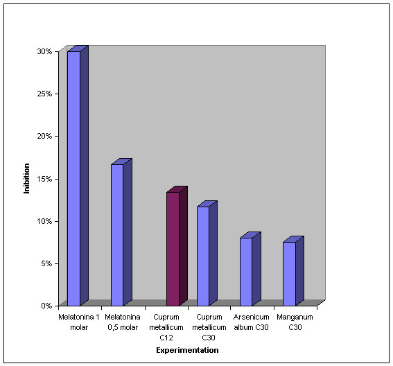
EXPERIMENTS
5.
DISCUSSIONThe research focused on the homeopathic medicine use in
order to measure the probable antioxidant effect of this medicine in rat’s
brain homogenate in the lipidic peroxidation comparatively to the
melatonin, because the melatonin has proved to have antioxidant action in
vitro.Hahnnemann, in chapter IV, which talks about the
pharmaco-dynamic of the substances, mentions: “We are not able to
find out this immaterial force that is latent in the close essence of the
medicine just with the reazon’s efforts. Just by the experience
(experimentation) we can clearly notice the phenomena it provokes when it
acts in a heallhy organism” (Pustiglione, 2001).As the homeopathic medicine experimentation always
occurs in vitro, following the same reasoning, we made the experimentation
in vitro.The homeopathic medicine is widely used in the whole
planet, despite of no knowing its real action mechanism, once such
mechanisms are stell no perfectly explained.The experimentation in vitro can complement the
knowlegde of the homeopathic medicine pathogenese itself, as well as its
action in the several organic alterations.This effect reproduction opens perspectives to other
essays that allow a better comprehension of the homeopathic medicine
action in vivo.In this work it was possible to verify the antioxidant
action of the homeopathic medicine: cuprum metallicum, arsenicum album,
zincum metallicum and manganum, comparatively to the melatonin, inhibiting
the lipoperoxidation over the young male rat’s brain homogenate.The melatonin presented significant action in the
lipidic peroxidation inhibition, that’s why it severed use a comparative
pattern of reference in the use of the homeopathic medicine mentioned.These homeopathic medicines choice was due to th fact
of them being part of the mitochondrial and cytoplasmic superoxide
dismutase. The arsenicum album was chosen because it is na important
polychrestus whose pathogenese fits to the profile of the stressed
individual psychically, that eventually can cause cellular oxidative
stress. These elements passed through trituration process before being
diluted, what means they suffered succussion.The work showed the homeopathic medicines cuprum
metallicum, manganum and arsenicum album are significant as inhibitor in
vitro of the lipidic peroxidation in rat brain homogenate, comparatively
to the melatonin in substanteal dose through the melondialdehyde dosage.Through the experiments analysis it was noticed, having
the melatonin 0,125 M as a reference, less inhibition of the lipidic
peroxidation. There was more inhibition of the lipoperoxidation with the
use of the homeopathic medicines cuprum metallicum, manganum and arsenicum
album in the 6ª, 12ª and 30ª centesimals. However, the same effect was
not observed to the Zincum metallicum.The significance gotten through the cuprum metallicum
and Manganum experimentation can be explained by them acting effectively
in substancial dose in the cytoplamisc and mitochondrial superoxide
dismutase respectively, maybe by the action of trace element.Yet, there is a question: what logic reasoning or
theoretical pattern must be elaborated to explain the lipidic peroxidation
inhibition in the 30ª centesimal, that supplant the Avogrado number?Stell observe that as the inhibition mentioned happened
also with medicine whose dilution supplants the Avogrado number, we can
infer the existance of antioxidant mechanisms stell inknown, that must be
discovered.Once verified the lipidic peroxidation inhibition, in
rats brain homogenate in vitro, gotten by the use of homeopathic medicine,
it’s inferred na antioxidant effect even bigger when used in vivo,
considering the whole symptomatic of the individual.This work considered only the lipidic peroxidation,
however, it is open the possibility of studying the antioxidant action of
the homeopathic medicine in the other phases of ROS cascades.In general the LPx is the result of unbalance between
pro and antioxidant favoring the first ones (Harata et al, 1983; Kawase et
al, 1989).The antioxidants are very important in the mechanism of
action of several toxins. The oxidants involvement in several diseases is
well established, however there isn’t unanimity about it ( Halliwell and
Gutteridge, 1989).Philogenetically and ontogenetically the organism
develped antioxidant defense mechanisms, making possible the adaptation of
superior organnisms to the aerobic environmente where the 3 enzymes
superoxide dismutase, catalase and glutathione peroxidase are fundamental
for the pro and antioxidant balance. The use of exogenous substances in
order to mantain this balance can highly contribute to the human health.The melatonin in substanteal dose showed it is a mighty
antioxidant inhibiting satisfactorily the lipidic peroxidation. The idea
of using melatonin in different concentrations was to give comparative
support to the homeopathic medicine, because the melatonin has efficient
antioxidant effect ( Halliwell and Gutteridge, 1989).The confrontation of the homeophatic medicine highly
diluted with substantial melatonin doses, as well as the description of
their respective pathogeneses, serves as subsidy not only for a better
understanding of their antioxidant effects but also to try to understand
the own pathogeneses of these metals, extending this comprehension to the
pathogeneses of the homeopathic medicine in general.About the lipidic peroxidation inhibition gotten
through homeopathic medicine experimentation we can infer that a thing
happens because of the oligoelements action of such medicaments, or even
through the proper homeopathic action described in the pathogeneses.The reactive oxygen species are involved with an
immense of physiological reactions, as well as in processes of diseases
pretty known nowadays.In front of such processes, the organism tries to adapt
to the environment conditions where it lives through the homeostasys,
denomination introduced to the Biology by the North American physiologist
Walter Bradfort Cannom, in 1916. The homeostasys is the living being
hereditary property of lasting long in time, maintouning the morphological
and functional balance of its cells and tissues. The homeostasys is
maintained by another hereditary property, the self-regulation (Maffei,
1978).The homeostasys and self-regulation of the genotype
constitute the organism adaptation and compensation mechanisms to the
external agents, influencing this way not only at the time of a disease
manifestation as well as in the way of evolution and therapeutic action
(Maffei, 1978).This adaptation happens mainly when we refer to the
oxygen paradox. The living beings evolved passing from the anaerobic to
the aerobic conditions, what happened thanks to the efficient antioxidant
system development, where the enzymes superoxide dismutase, glutathione
peroxidase and catalase play a fundamental role to the species
preservation and perpetuation whose metabolism is aerobic (Halliwell and
Gutteridge, 1985).As the homeopathic medicine probably acts recovering
the homeostasys, it is possible that it happens due to the antioxidant
action of this medicament in rats brain homogenate.Admitting the Homeopathy is based in the
experimentation in vivo, it was assumed the possibility of demonstrating
experimentally in vitro the antioxidant effect of the homeopathic
medicine.It is important to remember that long ago before Claude
Bernard, HaHnemann used the experimentation in vivo, establishing, this
way, the pathogenese (Tetau, 1980). On this perspective we can assure, by
analogy, the functionality of the experimentation in vitro.We start by the premise that the medicine is just one,
only being variable the techniques and the therapeutic methods used by
different schools.Einstein said:
“Every religion, every art, every science are
branches from the same tree where these aspirations Focus on the human
life ennolument, raising it over the spheres of the existence merely
material and leading the individual to freedom” ( Monteiro, 1982).A controlled study was developed in a demonstrative way
that aimed to demonstrate the of the experimental method in vitro.It was based on the presupposition that, if the
antioxidantion phenomenon happens isolated, in vitro, in the conditions
verified by anology, there is a reasonable probability of being reproduced
in na individual as a whole, once the mechanism for what its action
happens is known and evaluated.In order to delimit properly the study in such a
universe, the conceitual and operational definition of the used terms was
clarifild and it was also explained the focus over what the analysis was
made (Andrade, 1995).In this way, the study was concentrated on the lipidic
peroxidation, suggesting however, the need of a better comprehension of
the other oxidative chain phases, what would make possible a greater
property in the homeopathic medicine application in the several diseases
and organic changes.The study stell aimed to measure the homeopathic
medicine action in the immediate ambit of the experimental research, for a
better understanding of its use in the human health.This works opens perspectives for a better
comprehension of the homeopathic medicine action as antioxidant, as well
as its general action in the organism.It important to point ou that, because fo the
characteristics of the experiment it is impossible to elect the similimum
medicament, what is supposed to have a more effective antioxidant action
when administrated to the living being, filling the individual whole
symptomatic.
6.
CONCLUSIONSThe melatonin showed to be more efficient in the
lipidic peroxidation in rats brain homogenate in 1 molar, concentration,
coming next the melatonin 0,5 molar and the melatonin 0,25 molar.The lipoperoxidation inhibition with the use of the
homeopathic medicine occurred in a decreasing scale, like the following:
cuprum metallicum C12, Cuprum metallicum C30, Arsenicum album C30,
Manganum C30 and Arsenicum album C12.The other groups analysed did not present significant
differences.It was observed significative inhibition of the
lipoperoxidation with the use of homeopathic medicines over the Avogrado
number, such as the cuprum metallicum C30 and the arsenicum album C30.The homeopathic medicine produced significative
inhibition of lipoperoxidation comparatively to the melatonin, showing na
antioxidant action in vitro, deducing that, maybe, when administered
according to the Law of similars positive answers can happen to the
individual in this same sense.
7.
ABREVIATIONS AND SYMBOLSCN – Ion cyanide
CuZn SOD – Superoxide dismutase copper and zinc
dependentDNA – Desonyribonucleic acid
DOPA 3,4 – dihydroxyphenylalanine
DTPA – Diethylenetriamine-pentacetic acid
Ec SOD – Superoxide dismutase extracellular
ROS – Reactive Oxygen species
GPx – Glutathione peroxidase
GSH – Glutathione reduced
GSSG – Glutathione oxidated
Hb – Hemoglobin
HCN – Hydrogen cyanide; cyanidric acid
H2O2 – Hydrogen peroxide
HO· – Hydroxyl
KCN – Potassium cyanide
LPx – Lipoperoxidation
MDA – hyde
Mn SOD – Superoxide dismutase manganese dependent
NADP – Nicotinamide Adenine dinucleotide phosphate,
reduced formNADPH – Nicotinamide adenine dinucleotide phosphate,
oxidate formNBT – Blue of nitrotetrazole
O2 – Dioxygen; molecular oxygen
O2· –
Superoxide
1
O2 – Oxigênio Singlet
RNAm
–
Ribonucleic acid messagerRO· – Alcoxil radical
ROO· – Peroxil radical
ROOH – Organic Hydroperoxide
SOD – Superoxide dismutase
TBA – Tiobarbituric acid
TCA
– Trichloroacetic acid
8.
BIBLIOGRAPHIC REFERENCESAMES, B.N. – Dietary carcinogens and anticarcinogens
– oxyten radicals and degenerative diseases, Science, 221:1256, 1983AMES, B.N.& GOLD, L.S – Endogenous mutagens and
the causes of aging and cancer: Mutat. Res., 250:3-16, 1991.ANDRADE, M.M. – Como Preparar Trabalho para Cursos de
Pós – Graduação. São Paulo. Ed. Atlas AS, 1995, 118 p.BAROLLO, CR. Aos que se tratam pela Homeopatia. 4ª
edição. São Paulo. Editora Typus, 1989, 93 p.BAST, A; HAENEN, G.R.M.M.; DOELMAN, C.J.A – Oxidants
and antioxidants: state of the art. Am.J.Med., 91 ( suppl 3C):
2S-13S,1991.BEUTLER, E; WEST, C:BEUTLER, B. – Eletrophoretic
polymorphism of glutathione peroxidase. Hum. Gent, 38:163-169, 1974BRUNINI, C. Aforismos de Hipócrates, São Paulo. Typus
IBEHE. 1998, 173 p.CASTRO, D. Homeopatia e Profilaxia. São Paulo,
Editoração Mauro Familiar Editora. 1980, 145 p.CHADA, S; WHITNEY, C; NEWBURGER, PE –
Post-transcriptional regulation of glutathione peroxidase gene expression
by selenium in the HL-60 human myeloid cell line. Blood, 74 (7):
2535-2541, 1989.CHANCE, B- Effect of pH upon the reaction kinetics of
the enzyme-substrate compounds of catalase. J. Biol. Chem.,
194(2):471-481, 1952a.________ Effect of pH upon the reaction kinetics of the
enzyme-substrate copounds of catalase. J. Biol. Chem, 194 (2): 483-496,
1952b.CHANCE, B. & OSHINO, N – Dinetics and mechanisms
of catalase in peroxisomes of the mitochondrial fraction. Biochem J.,
122:225-233, 1971.CIRIOLO, M.R., FISKIN, K. DE MARTINO, A; CORASANITI,
M.T.; NISTICO, G; RTILIO G. – Age-related changes in Cu, Zn, superoxide
dismutase, Se-dependent and – independent glutathione peroxidase and
catalase activities in specific areas of rat brain. Ageing Dev.,
61:287-297, 1991.COHEN, G. & HOCHSTEIN, P. – Glutathione
peroxidase: the primary agent for the elimination of hydrogen peroxide in
erythrocytes. Biochemistry, 2(6): 1420-1428, 1963.CUTLER, R.G. – Evolution of human longevity and the
genetic complexity governing again rate. Proc.Natl.Acad. Sci. USA, 72(11):
4664-4668, 1975.________ Human longevity and againg: possible role of
reactive oxygen species. Ann NY Acad. Sci., 621:1-28, 1991.DEMARQUE, D – Técnicas Homeopáticas. Buenos Aires,
Ediciones Marecel, 1981, 328 p.DUPRAT, H . A teoria e a Técnica da Homeopatia, Rio de
Janeiro Gráfica Olímpica Editora, 1982, 198 p.EATON, J.W – Catalase and peroxidases and glutathione
na hydrogen peroxide: mysteries of the bestiary. J. Lab. Clin. Med.,
118:3-4, 1991FORSTRON, J W; ZAKOWSKI, JJ; TAPEL, A L. –
Identification of the catalytic site of rat liver glutathione peroxidase
as selenocysteine. Biochemistry, 17(13): 2639-2644, 1978FRIDOVICH, I – The biology of oxygen radicals.
Science, 201:875880. 1978.GAETANI, G.F.; GALIANO, S; CANEPA, L; FERRARIS, A M;
KIRKMAN, H N – Catalase and glutathione peroxidase are equally active in
detoxification of hydrogen peroxide in human erythrocytes. Blood, 73(1):
334-339, 1989.GERCHMAN, R; GILBER, D L ; NYE, S W; DWYER, P ; FENN W
O – Oxygen poisoning and X-irradiation: a mechanism in common. Science,
119:623-626, 1954GONZALEZ, M J – Lipid peroxidation and tumor grown:
na inverse relationship. Med Hypotheses, 38 (2): 106-110, 1992GUYTON, A C – Tratado de Fisiologia Médica. 4ª
edição. Rio de Janeiro. Ed Guanabara Hoogan S/A, 1973, 974 p.HALLIWELL, B. – Oxidants and human disease: some new
concepts. FASEB J, 1: 358-364, 1987._______ Oxidants and the central nervous system: some
fundamental questions. Acta Neurol. Scand., 126:23-33, 1989b._______ How to characterize a biological antioxidant.
Free rad. Res. Commun, 9 (1): 1-32, 1990._______ Reactive ozygen species in living systems:
source, biochemistry, and role in human disease. Am. J. med, 91 (suppl
3C): 14S-22S, 1991.HALLIWELL, B & GROTTLVELD, M. – The measurement
of free radical reactions in humans, FEBS Lett, 213(1):9-14, 1987.HALLIWELL B. & GUTTERIDGE, J M C – Free redicals
in Biology and Medicine. Oxford: Clerendon Press, 1985, 543 p.________ Free radicals in Biology and Medicine. Oxford:
Clarendon Press, 1989, 543 p.HALLIWELL , B; HOULT, R; BLAKE D R – Oxidants,
inflammation, and anti-inflammatory drugs. FASEB J, 2:2867-2873, 1988.HAHNEMANN, S. Matéria Médica Pura. 2ª edição. New
Delhi. Jain Publishing Co, 1920._________ Doenças Crônicas – Sua natureza Peculiar
e Sua Cura Homeopática. New Delhi. Jain Publishing Co, 1984HARMAN, D – The aging process. Proc. Natl. Acad. Sci.
USA, 78 (11): 7124-7128, 1981.HENDLER, SS – Vitamin and Mineral Encyclopedia. San
Diego, Simon e Schuski, 1990, 576 p.KAY, M M B; BOSMAN, G J C G M; SHAPIRO, S S ; BENDICH,
A ; BASSEL, P S – Oxidation as a possible mechanism of cellular aging:
Vitamin E deficiency causes premature aging and IgG binding to
erythrocytes. Proc. Natl. Acad. Sci. USA 83: 2463-2467, 1986.KENT, J T . Filosofia Homeopática, Buenos Aires.
Editorial Albatroz. 1980, 342 p.LATHOUD. Matéria Médica Homeopática. Buenos Aires.
Editorial Albatroz, 1980, 868 p.MACHLIN, L J & BENDICH, A – Free radical tissue
damage: protective role of antioxidant nutrients. FASEB J. 1:441-445,
1987.MAFFEI, W E. – Os fundamentos da Medicina. 2º
edição. São Paulo. Artes Médicas, 1978, 781 p.MANNERVIK, B – Glutathione peroxidase. Methods
Enzymol, 113:490-495, 1985.MARKLUND , S L. – Human copper-containing superoxide
dismutase of high molecular weight. Proc. Natl. Acad. Sci. USA,
79:7634-7638, 1982.MASTERS, C & CRANE D – Isozymes and the micro
organization of organellar structure and function. In: Isozymes:
Structure, Function, and Use in Biology and Medicine. Wilwy-Liss, Inc,
1990. P 101-122.McBRIDE, O W.; MITCHELL, A; LEE, B J; MULLENBACH, G;
HATFIELD, D – Gene for selenium-dependent glutathione peroxidase maps to
human chomosomes 3,21 and X BioFactors, 1 (4):285-292, 1988.McCORD, J M & FRIDOVICH, I – Superoxide dismutase
na enzymic function for erythrocuprein ( hemocuprein). J.Biol. Chem., 244
(22) 6049-6055, 1969.MEERA KHAN, P; VERMA, C; WIJNEN, L M M.; JAIRAJ, S –
Red cell glutathione peroxidase (GPX1) variation in Afro-Jamaican, Asiatic
Indian na Dutch populations: is the GPX1*2 alled of “Thomas”
variant na Afrincan marker? Hum. Genet., 66: 352-355, 1984.MENETRIER, J . A medicina das funções. São Paulo.
Organon, Bio. Press, 2000, 222 p.MILLS, G C. – The purification and propertiers fo
glutathione peroxidase of erythorcytes J Biol. Cheim., 234:502-506, 1959.MITROPOULOS, D – Oligoterapia. Italy, Editorium
Milano, 1994, 137 p.MONTEIRO, I – Reflexões Filosóficas. Ed. São
Paulo. Alvorada, 1982, 124 p.PAGLIA, D E & VALENTINE, W.N. – Studies on the
quantitative and qualitative characterization of erythrocyte glutathione
peroxidase. J. Lab. Clin. Med, 70(1): 158-169, 1967.PERCY, M E. – Catalase: na old enzyme with a new roe?
Can. J. Biochem. Cell Biol., 62:1006-1014, 1984.PORTA, E. Tissue lipoperoxidation and lipofuscin
accumulation as influenced by age, rype of dietary fat and levels of
vitamin E in rats. Advances in the Biosciences, 64:37-74, 1987._______ Role of oxidative damage in the again process.
In: CHOW, C.K., ed. Cellular Antioxidant Defense Mechanisms. Vol. III,
Boca raton, FL: CRC Press, 1988. P. 1-52.POZETTI, G.L. “Mecanismo de Ação de Medicamento
Homeopático”. São Paulo, Revista de Homeopatia, 1986, 66 p.PUSTIGLIONE, M & CARRILO JR., R. Organon da Arte de
Curar de Samuel Hahnemann – Versão para o português sistematizada e
comentada por Marcelo Pustiglione e Romeu Carrilo Jr. São Paulo,
Homeopatia Hoje, 1994, 205 p.REITER, J. R. & MAESTRONI, G J M. Melatonin in
relation to the antioxidative defense and immune systems: possible
implications for cell and organ transplantation, J. Mol. Med.,
77:36-39,1999.SCOTT, M.D.; LUBIN, B. H.; ZUO, L; KUYPERS, F A –
Erythrocyte defense against hydrogen peroxide: preeminent importance of
catalase. J. Lab. Clin. Med, 118:7-16, 1991.SIE, H – Oxidative stress: from basic research to
clinical application. Am. J.Med., 91 ( suppl 3C): 31S-38S, 1991.SINET, P.M ; MICHELSON, A M ; BAZIN, A; LEJEUNE, J ;
JÉROME, H – Increase in glutathione peroxidase activity in erythrocytes
from trisomy 21 subjects. Biochem. Biophys Res. Commun, 67 (3): 910-915,
1975.SINET, P M.; GARBER, P; JÉROME, H – Inativaction of
human CuZn superoxide dismutase during expossure to superoxide radical and
hydrogen peroxide Bull. Europ. Physiopath. Resp., 17 ( suppl.): 91-97,
1981SLATER, T.F – Free radical mechanism in tissue
injury. Biochem J, 222:1-15, 1984.SMITH, L; THIER, S.O – Fisiologia. 2ª ed. São
Paulo. Panamericana, 1990, 1260 p.SUN, Y & OBERLEY, L. W. – The inibition of
catalase by glutathione. Free Rad. Biol. Med, 7:595-602, 1989.TETAU, M – Homeopatia. 6º edição. São Paulo,
Andrei, 1980, 175 p.TORTI, A . Gli oligoelementi nel futuro terapêutico.
Milão. Giusepe Mario Ricchiuto Editore, 1988, 232 p.VANNIER, L . Compendio de Matéria Médica
Homeopática. Médico. Editorial Porrua. S. A 1979.VIJNOVSKY, B. Sintomas – Clave de La Materia Medica
Homeopatica – Buenos Aires – Editorial Albatros. 1978, 250 p.WEACKER, P; MUELLER, C.R; MAYEROVA, A; GRZESCHIK, K L;
ROPERS, H.H – Assignment of the gene coding for human catalase to the
short arm of chromosome 11. Ann. Genét., 23(2): 73-77, 1980.
Copyright
© CELSO FERNANDES BATELLO 2003